 Movie
Movie Controller

[English] 日本語
 Yorodumi
- PDB-3daa: CRYSTALLOGRAPHIC STRUCTURE OF D-AMINO ACID AMINOTRANSFERASE INACT... -
+ Open data

- Basic information
| Entry | Database: PDB / ID: 3daa | ||||||
|---|---|---|---|---|---|---|---|
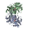
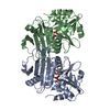
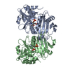
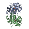
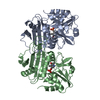
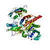
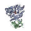
| Title | CRYSTALLOGRAPHIC STRUCTURE OF D-AMINO ACID AMINOTRANSFERASE INACTIVATED BY PYRIDOXYL-D-ALANINE | ||||||
 Components Components | D-AMINO ACID AMINOTRANSFERASE | ||||||
 Keywords Keywords | AMINOTRANSFERASE / PYRIDOXAL PHOSPHATE / TRANSAMINASE | ||||||
| Function / homology |  Function and homology information Function and homology informationD-amino acid biosynthetic process / D-amino-acid transaminase / D-alanine:2-oxoglutarate transaminase activity / D-amino acid catabolic process / pyridoxal phosphate binding / cytosol Similarity search - Function | ||||||
| Biological species |  | ||||||
| Method |  X-RAY DIFFRACTION / SYNCHROTRON / MOLECULAR REPLACEMENT / Resolution: 1.9 Å X-RAY DIFFRACTION / SYNCHROTRON / MOLECULAR REPLACEMENT / Resolution: 1.9 Å | ||||||
 Authors Authors | Peisach, D. / Chipman, D.M. / Ringe, D. | ||||||
 Citation Citation | Journal: Biochemistry / Year: 1998 Title: Crystallographic study of steps along the reaction pathway of D-amino acid aminotransferase. Authors: Peisach, D. / Chipman, D.M. / Van Ophem, P.W. / Manning, J.M. / Ringe, D. #1: Journal: Biochemistry / Year: 1995Title: Crystal Structure of a D-Amino Acid Aminotransferase: How the Protein Controls Stereoselectivity Authors: Sugio, S. / Petsko, G.A. / Manning, J.M. / Soda, K. / Ringe, D. | ||||||
| History |
|
- Structure visualization
| Structure viewer | Molecule: MolmilJmol/JSmol |
|---|
- Downloads & links
-Download
| PDBx/mmCIF format | 3daa.cif.gz | 125.8 KB | Display | PDBx/mmCIF format |
|---|---|---|---|---|
| PDB format | pdb3daa.ent.gz | 98.3 KB | Display | PDB format |
| PDBx/mmJSON format | 3daa.json.gz | Tree view | PDBx/mmJSON format | |
| Others |  Other downloads Other downloads |
-Validation report
| Arichive directory | https://data.pdbj.org/pub/pdb/validation_reports/da/3daaftp://data.pdbj.org/pub/pdb/validation_reports/da/3daa | HTTPS FTP |
|---|
-Related structure data
| Related structure data |  4daaC  1daaS S: Starting model for refinement C: citing same article ( |
|---|---|
| Similar structure data |


-Links
 PDBj
PDBj
- Assembly
| Deposited unit | 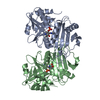
| ||||||||
|---|---|---|---|---|---|---|---|---|---|
| 1 |
| ||||||||
| Unit cell |
|
-Components
| #1: Protein | Mass: 31721.156 Da / Num. of mol.: 2 Source method: isolated from a genetically manipulated source Source: (gene. exp.) #2: Chemical |   Type: D-peptide linking / Mass: 320.236 Da / Num. of mol.: 2 / Source method: obtained synthetically / Formula: C11H17N2O7P Type: D-peptide linking / Mass: 320.236 Da / Num. of mol.: 2 / Source method: obtained synthetically / Formula: C11H17N2O7P#3: Water | ChemComp-HOH / |  Mass: 18.015 Da / Num. of mol.: 253 / Source method: isolated from a natural source / Formula: H2O Mass: 18.015 Da / Num. of mol.: 253 / Source method: isolated from a natural source / Formula: H2O |
|---|
-Experimental details
-Experiment
| Experiment | Method: X-RAY DIFFRACTION / Number of used crystals: 1 |
|---|
- Sample preparation
| Crystal | Density Matthews: 2.1 Å3/Da / Density % sol: 42 % | |||||||||||||||||||||||||||||||||||||||||||||||||||||||
|---|---|---|---|---|---|---|---|---|---|---|---|---|---|---|---|---|---|---|---|---|---|---|---|---|---|---|---|---|---|---|---|---|---|---|---|---|---|---|---|---|---|---|---|---|---|---|---|---|---|---|---|---|---|---|---|---|
| Crystal grow | Method: vapor diffusion, hanging drop / pH: 8 Details: PDA INACTIVATED PROTEIN WAS CONCENTRATED TO 30 MG/ML IN 0.1 M POTASSIUM PHOSPHATE BUFFER PH 7.6 CONTAINING 50 UM PLP AND 0.001 BETA-MERCAPTOETHANOL. CRYSTALS WERE THEN GROWN BY THE HANGING ...Details: PDA INACTIVATED PROTEIN WAS CONCENTRATED TO 30 MG/ML IN 0.1 M POTASSIUM PHOSPHATE BUFFER PH 7.6 CONTAINING 50 UM PLP AND 0.001 BETA-MERCAPTOETHANOL. CRYSTALS WERE THEN GROWN BY THE HANGING DROP METHOD IN 27% PEG 4000, 0.3 M SODIUM ACETATE, AND 0.1 M TRIS-CHLORIDE PH 8.5., vapor diffusion - hanging drop PH range: 7.6-8.5 | |||||||||||||||||||||||||||||||||||||||||||||||||||||||
| Crystal | *PLUS | |||||||||||||||||||||||||||||||||||||||||||||||||||||||
| Crystal grow | *PLUS pH: 7.6 / Method: vapor diffusion, hanging drop | |||||||||||||||||||||||||||||||||||||||||||||||||||||||
| Components of the solutions | *PLUS
|
-Data collection
| Diffraction | Mean temperature: 100 K |
|---|---|
| Diffraction source | Source: SYNCHROTRON / Site: NSLS  / Beamline: X12C / Wavelength: 0.97 / Beamline: X12C / Wavelength: 0.97 |
| Detector | Type: MARRESEARCH / Detector: IMAGE PLATE / Date: Jun 14, 1996 / Details: MIRROR |
| Radiation | Monochromator: FLAT CRYSTAL / Monochromatic (M) / Laue (L): M / Scattering type: x-ray |
| Radiation wavelength | Wavelength: 0.97 Å / Relative weight: 1 |
| Reflection | Resolution: 1.8→30 Å / Num. obs: 43763 / % possible obs: 87.1 % / Observed criterion σ(I): 0 / Redundancy: 2.47 % / Rmerge(I) obs: 0.062 / Net I/σ(I): 15.9 |
| Reflection shell | Resolution: 1.8→1.85 Å / Redundancy: 2.35 % / Rmerge(I) obs: 0.293 / Mean I/σ(I) obs: 3.5 / % possible all: 76.1 |
| Reflection | *PLUS Num. measured all: 57553 |
| Reflection shell | *PLUS % possible obs: 76.1 % |
- Processing
| Software |
| ||||||||||||||||||||||||||||||||||||||||||||||||||||||||||||
|---|---|---|---|---|---|---|---|---|---|---|---|---|---|---|---|---|---|---|---|---|---|---|---|---|---|---|---|---|---|---|---|---|---|---|---|---|---|---|---|---|---|---|---|---|---|---|---|---|---|---|---|---|---|---|---|---|---|---|---|---|---|
| Refinement | Method to determine structure: MOLECULAR REPLACEMENT Starting model: PDB ENTRY 1DAA Resolution: 1.9→30 Å / Rfactor Rfree error: 0.005 / Data cutoff high absF: 10000000 / Data cutoff low absF: 0.001 / Cross valid method: THROUGHOUT / σ(F): 0 / Details: USED SOLVENT MASK DURING REFINEMENT
| ||||||||||||||||||||||||||||||||||||||||||||||||||||||||||||
| Refine analyze | Luzzati d res low obs: 30 Å | ||||||||||||||||||||||||||||||||||||||||||||||||||||||||||||
| Refinement step | Cycle: LAST / Resolution: 1.9→30 Å
| ||||||||||||||||||||||||||||||||||||||||||||||||||||||||||||
| Refine LS restraints |
| ||||||||||||||||||||||||||||||||||||||||||||||||||||||||||||
| LS refinement shell | Resolution: 1.9→1.99 Å / Rfactor Rfree error: 0.023 / Total num. of bins used: 8
| ||||||||||||||||||||||||||||||||||||||||||||||||||||||||||||
| Xplor file |
| ||||||||||||||||||||||||||||||||||||||||||||||||||||||||||||
| Software | *PLUS Name: X-PLOR / Version: 3.8 / Classification: refinement | ||||||||||||||||||||||||||||||||||||||||||||||||||||||||||||
| Refine LS restraints | *PLUS
|
