 Movie
Movie Controller

[English] 日本語
 Yorodumi
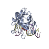
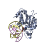
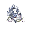
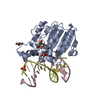
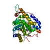
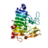
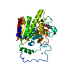
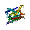
- PDB-1f6o: CRYSTAL STRUCTURE OF THE HUMAN AAG DNA REPAIR GLYCOSYLASE COMPLEX... -
+ Open data

- Basic information
| Entry | Database: PDB / ID: 1f6o | ||||||
|---|---|---|---|---|---|---|---|
| Title | CRYSTAL STRUCTURE OF THE HUMAN AAG DNA REPAIR GLYCOSYLASE COMPLEXED WITH DNA | ||||||
 Components Components |
| ||||||
 Keywords Keywords | HYDROLASE/DNA / Protein-DNA Complex / AAG DNA repair glycosylase / HYDROLASE-DNA COMPLEX | ||||||
| Function / homology |  Function and homology information Function and homology informationalkylbase DNA N-glycosylase activity / DNA-3-methyladenine glycosylase II / depurination / DNA N-glycosylase activity / Displacement of DNA glycosylase by APEX1 / DNA alkylation repair / mitochondrial nucleoid / Recognition and association of DNA glycosylase with site containing an affected purine / Cleavage of the damaged purine / base-excision repair ...alkylbase DNA N-glycosylase activity / DNA-3-methyladenine glycosylase II / depurination / DNA N-glycosylase activity / Displacement of DNA glycosylase by APEX1 / DNA alkylation repair / mitochondrial nucleoid / Recognition and association of DNA glycosylase with site containing an affected purine / Cleavage of the damaged purine / base-excision repair / damaged DNA binding / mitochondrion / DNA binding / nucleoplasm / cytosol Similarity search - Function | ||||||
| Biological species |  Homo sapiens (human) Homo sapiens (human) | ||||||
| Method |  X-RAY DIFFRACTION / SYNCHROTRON / Resolution: 2.4 Å X-RAY DIFFRACTION / SYNCHROTRON / Resolution: 2.4 Å | ||||||
 Authors Authors | Lau, A.Y. / Wyatt, M.D. / Glassner, B.J. / Samson, L.D. / Ellenberger, T. | ||||||
 Citation Citation | Journal: Proc.Natl.Acad.Sci.USA / Year: 2000 Title: Molecular basis for discriminating between normal and damaged bases by the human alkyladenine glycosylase, AAG. Authors: Lau, A.Y. / Wyatt, M.D. / Glassner, B.J. / Samson, L.D. / Ellenberger, T. | ||||||
| History |
|
- Structure visualization
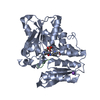
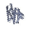
| Structure viewer | Molecule: MolmilJmol/JSmol |
|---|
- Downloads & links
-Download
| PDBx/mmCIF format | 1f6o.cif.gz | 73.8 KB | Display | PDBx/mmCIF format |
|---|---|---|---|---|
| PDB format | pdb1f6o.ent.gz | 51 KB | Display | PDB format |
| PDBx/mmJSON format | 1f6o.json.gz | Tree view | PDBx/mmJSON format | |
| Others |  Other downloads Other downloads |
-Validation report
| Arichive directory | https://data.pdbj.org/pub/pdb/validation_reports/f6/1f6oftp://data.pdbj.org/pub/pdb/validation_reports/f6/1f6o | HTTPS FTP |
|---|
-Related structure data
-Links
 PDBj
PDBj


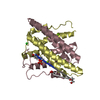
- Assembly
| Deposited unit | 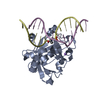
| ||||||||||
|---|---|---|---|---|---|---|---|---|---|---|---|
| 1 |
| ||||||||||
| Unit cell |
|
-Components
| #1: DNA chain | Mass: 3832.501 Da / Num. of mol.: 1 / Source method: obtained synthetically |
|---|---|
| #2: DNA chain | Mass: 3975.611 Da / Num. of mol.: 1 / Source method: obtained synthetically |
| #3: Protein | Mass: 24328.916 Da / Num. of mol.: 1 / Fragment: C-TERMINAL FRAGMENT Source method: isolated from a genetically manipulated source Source: (gene. exp.) Homo sapiens (human) / Plasmid: PLM1 / Production host:  References: UniProt: P29372, DNA-3-methyladenine glycosylase I |
| #4: Chemical | ChemComp-NA /   Mass: 22.990 Da / Num. of mol.: 1 / Source method: obtained synthetically / Formula: Na Mass: 22.990 Da / Num. of mol.: 1 / Source method: obtained synthetically / Formula: Na |
| #5: Water | ChemComp-HOH /  Mass: 18.015 Da / Num. of mol.: 134 / Source method: isolated from a natural source / Formula: H2O Mass: 18.015 Da / Num. of mol.: 134 / Source method: isolated from a natural source / Formula: H2O |
-Experimental details
-Experiment
| Experiment | Method: X-RAY DIFFRACTION / Number of used crystals: 1 |
|---|
- Sample preparation
| Crystal | Density Matthews: 2.47 Å3/Da / Density % sol: 50.27 % | ||||||||||||||||||||||||||||||
|---|---|---|---|---|---|---|---|---|---|---|---|---|---|---|---|---|---|---|---|---|---|---|---|---|---|---|---|---|---|---|---|
| Crystal grow | Temperature: 298 K / Method: vapor diffusion, hanging drop / pH: 6.5 Details: PEG 8000, magnesium acetate, sodium cacodylate, pH 6.5, VAPOR DIFFUSION, HANGING DROP, temperature 298K | ||||||||||||||||||||||||||||||
| Components of the solutions |
| ||||||||||||||||||||||||||||||
| Crystal grow | *PLUS pH: 7.5 / Method: unknown | ||||||||||||||||||||||||||||||
| Components of the solutions | *PLUS
|
-Data collection
| Diffraction | Mean temperature: 100 K |
|---|---|
| Diffraction source | Source: SYNCHROTRON / Site: NSLS  / Beamline: X12C / Wavelength: 1 / Beamline: X12C / Wavelength: 1 |
| Detector | Type: PHILLIPS / Detector: CCD / Date: Jun 6, 1999 |
| Radiation | Protocol: SINGLE WAVELENGTH / Monochromatic (M) / Laue (L): M / Scattering type: x-ray |
| Radiation wavelength | Wavelength: 1 Å / Relative weight: 1 |
| Reflection | Resolution: 2.4→500 Å / Num. all: 13118 / Num. obs: 11332 / % possible obs: 86.4 % / Observed criterion σ(I): 2 / Redundancy: 8 % / Biso Wilson estimate: 41.8 Å2 / Rmerge(I) obs: 0.071 / Net I/σ(I): 21.5 |
| Reflection shell | Resolution: 2.4→2.53 Å / Redundancy: 6 % / Rmerge(I) obs: 0.253 / Num. unique all: 1643 / % possible all: 75.6 |
| Reflection | *PLUS Num. obs: 13252 / % possible obs: 86.1 % / Num. measured all: 100868 |
| Reflection shell | *PLUS % possible obs: 75.6 % / Mean I/σ(I) obs: 3.5 |
- Processing
| Software |
| |||||||||||||||||||||||||
|---|---|---|---|---|---|---|---|---|---|---|---|---|---|---|---|---|---|---|---|---|---|---|---|---|---|---|
| Refinement | Resolution: 2.4→500 Å / Cross valid method: THROUGHOUT / σ(F): 4 / σ(I): 2 / Stereochemistry target values: Engh & Huber Details: Used Powell conjugate gradient minimization and torsion angle-restrained molecular dynamics
| |||||||||||||||||||||||||
| Refinement step | Cycle: LAST / Resolution: 2.4→500 Å
| |||||||||||||||||||||||||
| Refine LS restraints |
|
