 Movie
Movie Controller

[English] 日本語
 Yorodumi
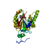
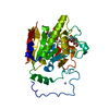
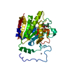
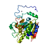
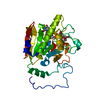
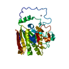
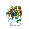
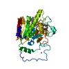
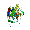
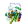
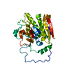
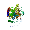
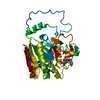
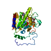
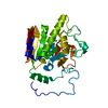
- PDB-1zi3: Crystal Structure of Human N-acetylgalactosaminyltransferase (GTA... -
+ Open data

- Basic information
| Entry | Database: PDB / ID: 1zi3 | |||||||||
|---|---|---|---|---|---|---|---|---|---|---|
| Title | Crystal Structure of Human N-acetylgalactosaminyltransferase (GTA) Complexed with N-acetyllactosamine | |||||||||
 Components Components | Histo-blood group ABO system transferase (NAGAT) Includes: Glycoprotein-fucosylgalactoside alpha-N- acetylgalactosaminyltransferase | |||||||||
 Keywords Keywords | TRANSFERASE / GTA / ABO(H) / H-antigen / blood groups / glycosyltransferase | |||||||||
| Function / homology |  Function and homology information Function and homology informationfucosylgalactoside 3-alpha-galactosyltransferase / glycoprotein-fucosylgalactoside alpha-N-acetylgalactosaminyltransferase / glycoprotein-fucosylgalactoside alpha-N-acetylgalactosaminyltransferase activity / fucosylgalactoside 3-alpha-galactosyltransferase activity / ABO blood group biosynthesis / Golgi cisterna membrane / antigen binding / manganese ion binding / carbohydrate metabolic process / vesicle ...fucosylgalactoside 3-alpha-galactosyltransferase / glycoprotein-fucosylgalactoside alpha-N-acetylgalactosaminyltransferase / glycoprotein-fucosylgalactoside alpha-N-acetylgalactosaminyltransferase activity / fucosylgalactoside 3-alpha-galactosyltransferase activity / ABO blood group biosynthesis / Golgi cisterna membrane / antigen binding / manganese ion binding / carbohydrate metabolic process / vesicle / Golgi membrane / nucleotide binding / Golgi apparatus / extracellular region Similarity search - Function | |||||||||
| Biological species |  Homo sapiens (human) Homo sapiens (human) | |||||||||
| Method |  X-RAY DIFFRACTION / MOLECULAR REPLACEMENT / Resolution: 1.69 Å X-RAY DIFFRACTION / MOLECULAR REPLACEMENT / Resolution: 1.69 Å | |||||||||
 Authors Authors | Letts, J.A. / Rose, N.L. / Fang, Y.R. / Barry, C.H. / Borisova, S.N. / Seto, N.O. / Palcic, M.M. / Evans, S.V. | |||||||||
 Citation Citation | Journal: J.Biol.Chem. / Year: 2006 Title: Differential Recognition of the Type I and II H Antigen Acceptors by the Human ABO(H) Blood Group A and B Glycosyltransferases. Authors: Letts, J.A. / Rose, N.L. / Fang, Y.R. / Barry, C.H. / Borisova, S.N. / Seto, N.O. / Palcic, M.M. / Evans, S.V. | |||||||||
| History |
|
- Structure visualization
| Structure viewer | Molecule: MolmilJmol/JSmol |
|---|
- Downloads & links
-Download
| PDBx/mmCIF format | 1zi3.cif.gz | 74.6 KB | Display | PDBx/mmCIF format |
|---|---|---|---|---|
| PDB format | pdb1zi3.ent.gz | 53.1 KB | Display | PDB format |
| PDBx/mmJSON format | 1zi3.json.gz | Tree view | PDBx/mmJSON format | |
| Others |  Other downloads Other downloads |
-Validation report
| Arichive directory | https://data.pdbj.org/pub/pdb/validation_reports/zi/1zi3ftp://data.pdbj.org/pub/pdb/validation_reports/zi/1zi3 | HTTPS FTP |
|---|
-Related structure data
| Related structure data | 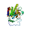 1zhjC 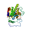 1zi1C  1zi4C  1zi5C 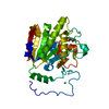 1zizC 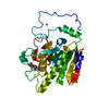 1zj0C 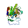 1zj1C 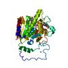 1zj2C  1zj3C  1zjoC 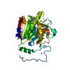 1zjpC 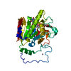 2a8uC  2a8wC 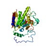 1lz0S C: citing same article ( S: Starting model for refinement |
|---|---|
| Similar structure data |


-Links
 PDBj
PDBj


- Assembly
| Deposited unit | 
| ||||||||
|---|---|---|---|---|---|---|---|---|---|
| 1 |
| ||||||||
| 2 | 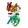
| ||||||||
| Unit cell |
|
-Components
| #1: Protein | Mass: 34194.496 Da / Num. of mol.: 1 Source method: isolated from a genetically manipulated source Source: (gene. exp.) Homo sapiens (human) / Gene: ABO / Plasmid: PCW DELTA 1AC (E-H) / Production host:  References: UniProt: P16442, glycoprotein-fucosylgalactoside alpha-N-acetylgalactosaminyltransferase | ||||
|---|---|---|---|---|---|
| #2: Polysaccharide | beta-D-galactopyranose-(1-4)-2-acetamido-2-deoxy-alpha-D-glucopyranose / N-acetyl-alpha-lactosamine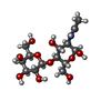  Source method: isolated from a genetically manipulated source Details: oligosaccharide / References: N-acetyl-alpha-lactosamine | ||||
| #3: Chemical | ChemComp-HG /   Mass: 200.590 Da / Num. of mol.: 4 / Source method: obtained synthetically / Formula: Hg Mass: 200.590 Da / Num. of mol.: 4 / Source method: obtained synthetically / Formula: Hg#4: Chemical | ChemComp-CL / |   Mass: 35.453 Da / Num. of mol.: 1 / Source method: obtained synthetically / Formula: Cl Mass: 35.453 Da / Num. of mol.: 1 / Source method: obtained synthetically / Formula: Cl#5: Water | ChemComp-HOH / |  Mass: 18.015 Da / Num. of mol.: 200 / Source method: isolated from a natural source / Formula: H2O Mass: 18.015 Da / Num. of mol.: 200 / Source method: isolated from a natural source / Formula: H2O |
-Experimental details
-Experiment
| Experiment | Method: X-RAY DIFFRACTION / Number of used crystals: 1 |
|---|
- Sample preparation
| Crystal | Density Matthews: 2.27 Å3/Da / Density % sol: 45.84 % |
|---|
-Data collection
| Diffraction | Mean temperature: 113 K |
|---|---|
| Detector | Type: RIGAKU RAXIS IV / Detector: IMAGE PLATE / Date: Feb 18, 2005 |
| Radiation | Protocol: SINGLE WAVELENGTH / Monochromatic (M) / Laue (L): M / Scattering type: x-ray |
| Radiation wavelength | Relative weight: 1 |
- Processing
| Software |
| ||||||||||||||||||
|---|---|---|---|---|---|---|---|---|---|---|---|---|---|---|---|---|---|---|---|
| Refinement | Method to determine structure: MOLECULAR REPLACEMENT Starting model: 1LZ0 Resolution: 1.69→20 Å /
| ||||||||||||||||||
| Refinement step | Cycle: LAST / Resolution: 1.69→20 Å
|
