 Movie
Movie Controller

[English] 日本語
 Yorodumi
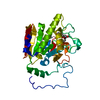
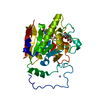
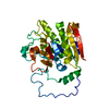
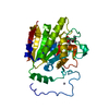
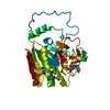
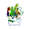
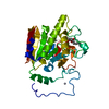
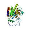
- PDB-1r7v: Glycosyltransferase A in complex with 3-amino-acceptor analog inh... -
+ Open data

- Basic information
| Entry | Database: PDB / ID: 1r7v | |||||||||
|---|---|---|---|---|---|---|---|---|---|---|
| Title | Glycosyltransferase A in complex with 3-amino-acceptor analog inhibitor | |||||||||
 Components Components | Glycoprotein-fucosylgalactoside alpha-N-acetylgalactosaminyltransferase | |||||||||
 Keywords Keywords | TRANSFERASE / Glycoprotein / transmembrane / signal-anchor / blood group antigen | |||||||||
| Function / homology |  Function and homology information Function and homology informationfucosylgalactoside 3-alpha-galactosyltransferase / glycoprotein-fucosylgalactoside alpha-N-acetylgalactosaminyltransferase / glycoprotein-fucosylgalactoside alpha-N-acetylgalactosaminyltransferase activity / fucosylgalactoside 3-alpha-galactosyltransferase activity / ABO blood group biosynthesis / Golgi cisterna membrane / antigen binding / manganese ion binding / carbohydrate metabolic process / vesicle ...fucosylgalactoside 3-alpha-galactosyltransferase / glycoprotein-fucosylgalactoside alpha-N-acetylgalactosaminyltransferase / glycoprotein-fucosylgalactoside alpha-N-acetylgalactosaminyltransferase activity / fucosylgalactoside 3-alpha-galactosyltransferase activity / ABO blood group biosynthesis / Golgi cisterna membrane / antigen binding / manganese ion binding / carbohydrate metabolic process / vesicle / Golgi membrane / nucleotide binding / Golgi apparatus / extracellular region Similarity search - Function | |||||||||
| Biological species |  Homo sapiens (human) Homo sapiens (human) | |||||||||
| Method |  X-RAY DIFFRACTION / MOLECULAR REPLACEMENT / Resolution: 2.09 Å X-RAY DIFFRACTION / MOLECULAR REPLACEMENT / Resolution: 2.09 Å | |||||||||
 Authors Authors | Nguyen, H.P. / Seto, N.O.L. / Cai, Y. / Leinala, E.K. / Borisova, S.N. / Palcic, M.M. / Evans, S.V. | |||||||||
 Citation Citation | Journal: J.Biol.Chem. / Year: 2003 Title: The influence of an intramolecular hydrogen bond in differential recognition of inhibitory acceptor analogs by human ABO(H) blood group A and B glycosyltransferases Authors: Nguyen, H.P. / Seto, N.O.L. / Cai, Y. / Leinala, E.K. / Borisova, S.N. / Palcic, M.M. / Evans, S.V. #1: Journal: Acta Crystallogr.,Sect.D / Year: 1998Title: Crystallography & NMR System Authors: Brunger, A.T. / Adams, P.D. / Clore, G.M. / Delano, W.L. / Gros, P. / Grosse-Kunstleve, R. / Jiang, J.-S. / Kuszewski, J. / Nilges, M. / Pannu, N.S. / Read, R.J. / Rice, L.M. / Simonson, T. / Warren, G. | |||||||||
| History |
|
- Structure visualization
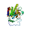
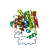
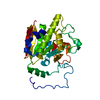
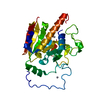
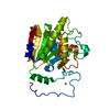
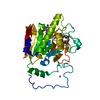
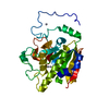
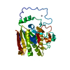
| Structure viewer | Molecule: MolmilJmol/JSmol |
|---|
- Downloads & links
-Download
| PDBx/mmCIF format | 1r7v.cif.gz | 74 KB | Display | PDBx/mmCIF format |
|---|---|---|---|---|
| PDB format | pdb1r7v.ent.gz | 53.1 KB | Display | PDB format |
| PDBx/mmJSON format | 1r7v.json.gz | Tree view | PDBx/mmJSON format | |
| Others |  Other downloads Other downloads |
-Validation report
| Arichive directory | https://data.pdbj.org/pub/pdb/validation_reports/r7/1r7vftp://data.pdbj.org/pub/pdb/validation_reports/r7/1r7v | HTTPS FTP |
|---|
-Related structure data
| Related structure data |  1r7tC  1r7uC  1r7xC 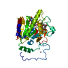 1r7yC 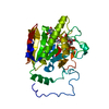 1r80C 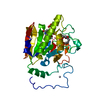 1r81C 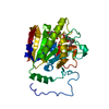 1r82C C: citing same article ( |
|---|---|
| Similar structure data |


-Links
 PDBj
PDBj


- Assembly
| Deposited unit | 
| ||||||||
|---|---|---|---|---|---|---|---|---|---|
| 1 |
| ||||||||
| 2 | 
| ||||||||
| Unit cell |
|
-Components
| #1: Protein | Mass: 32999.148 Da / Num. of mol.: 1 / Fragment: Catalytic Domain (Residues 63-345) Source method: isolated from a genetically manipulated source Source: (gene. exp.) Homo sapiens (human) / Production host:  References: UniProt: P16442, glycoprotein-fucosylgalactoside alpha-N-acetylgalactosaminyltransferase | ||
|---|---|---|---|
| #2: Polysaccharide | alpha-L-fucopyranose-(1-2)-hexyl 3-amino-3-deoxy-beta-D-galactopyranoside Type: oligosaccharide / Mass: 409.472 Da / Num. of mol.: 1 Source method: isolated from a genetically manipulated source | ||
| #3: Chemical | ChemComp-HG /   Mass: 200.590 Da / Num. of mol.: 4 / Source method: obtained synthetically / Formula: Hg Mass: 200.590 Da / Num. of mol.: 4 / Source method: obtained synthetically / Formula: Hg#4: Water | ChemComp-HOH / |  Mass: 18.015 Da / Num. of mol.: 155 / Source method: isolated from a natural source / Formula: H2O Mass: 18.015 Da / Num. of mol.: 155 / Source method: isolated from a natural source / Formula: H2O |
-Experimental details
-Experiment
| Experiment | Method: X-RAY DIFFRACTION / Number of used crystals: 1 |
|---|
- Sample preparation
| Crystal | Density Matthews: 2.42 Å3/Da / Density % sol: 48.67 % | ||||||||||||||||||||||||||||||||||||||||||||||||||||||||||||||||||||||||||||||||||||||||||||||||||||||||||||||||
|---|---|---|---|---|---|---|---|---|---|---|---|---|---|---|---|---|---|---|---|---|---|---|---|---|---|---|---|---|---|---|---|---|---|---|---|---|---|---|---|---|---|---|---|---|---|---|---|---|---|---|---|---|---|---|---|---|---|---|---|---|---|---|---|---|---|---|---|---|---|---|---|---|---|---|---|---|---|---|---|---|---|---|---|---|---|---|---|---|---|---|---|---|---|---|---|---|---|---|---|---|---|---|---|---|---|---|---|---|---|---|---|---|---|
| Crystal grow | Method: vapor diffusion, hanging drop / Details: VAPOR DIFFUSION, HANGING DROP | ||||||||||||||||||||||||||||||||||||||||||||||||||||||||||||||||||||||||||||||||||||||||||||||||||||||||||||||||
| Crystal grow | *PLUS Temperature: 18 ℃ / pH: 7.5 / Method: vapor diffusion, hanging dropDetails: Patenaude, S.I., (2002) Nat. Struct. Biol., 9, 685. | ||||||||||||||||||||||||||||||||||||||||||||||||||||||||||||||||||||||||||||||||||||||||||||||||||||||||||||||||
| Components of the solutions | *PLUS
|
-Data collection
| Diffraction | Mean temperature: 100 K |
|---|---|
| Diffraction source | Source: ROTATING ANODE / Type: RIGAKU RU300 / Wavelength: 1.5418 |
| Detector | Type: MARRESEARCH / Detector: AREA DETECTOR |
| Radiation | Protocol: SINGLE WAVELENGTH / Monochromatic (M) / Laue (L): M / Scattering type: x-ray |
| Radiation wavelength | Wavelength: 1.5418 Å / Relative weight: 1 |
| Reflection | Resolution: 2.09→19.32 Å / Num. all: 33375 / Observed criterion σ(F): 0 / Biso Wilson estimate: 25.2 Å2 / Limit h max: 24 / Limit h min: 0 / Limit k max: 71 / Limit k min: 0 / Limit l max: 38 / Limit l min: -38 / Observed criterion F max: 927674.87 / Observed criterion F min: 0.32 |
| Reflection | *PLUS Lowest resolution: 20 Å / Num. obs: 18560 / % possible obs: 96.6 % / Observed criterion σ(F): 0 / Rmerge(I) obs: 0.046 |
| Reflection shell | *PLUS % possible obs: 91.9 % / Rmerge(I) obs: 0.376 |
- Processing
| Software |
| ||||||||||||||||||||||||||||||||||||||||||||||||||||||||||||||||||||||||||||||||||||||||||||||||||||||||||||||
|---|---|---|---|---|---|---|---|---|---|---|---|---|---|---|---|---|---|---|---|---|---|---|---|---|---|---|---|---|---|---|---|---|---|---|---|---|---|---|---|---|---|---|---|---|---|---|---|---|---|---|---|---|---|---|---|---|---|---|---|---|---|---|---|---|---|---|---|---|---|---|---|---|---|---|---|---|---|---|---|---|---|---|---|---|---|---|---|---|---|---|---|---|---|---|---|---|---|---|---|---|---|---|---|---|---|---|---|---|---|---|---|
| Refinement | Method to determine structure: MOLECULAR REPLACEMENT / Resolution: 2.09→19.32 Å / Rfactor Rfree error: 0.004 / Occupancy max: 1 / Occupancy min: 0.5 / Cross valid method: THROUGHOUT
| ||||||||||||||||||||||||||||||||||||||||||||||||||||||||||||||||||||||||||||||||||||||||||||||||||||||||||||||
| Solvent computation | Solvent model: CNS bulk solvent model used / Bsol: 81.5588 Å2 / ksol: 0.412726 e/Å3 | ||||||||||||||||||||||||||||||||||||||||||||||||||||||||||||||||||||||||||||||||||||||||||||||||||||||||||||||
| Displacement parameters | Biso max: 129.09 Å2 / Biso mean: 42.16 Å2 / Biso min: 15.56 Å2
| ||||||||||||||||||||||||||||||||||||||||||||||||||||||||||||||||||||||||||||||||||||||||||||||||||||||||||||||
| Refine analyze |
| ||||||||||||||||||||||||||||||||||||||||||||||||||||||||||||||||||||||||||||||||||||||||||||||||||||||||||||||
| Refinement step | Cycle: LAST / Resolution: 2.09→19.32 Å
| ||||||||||||||||||||||||||||||||||||||||||||||||||||||||||||||||||||||||||||||||||||||||||||||||||||||||||||||
| Refine LS restraints |
| ||||||||||||||||||||||||||||||||||||||||||||||||||||||||||||||||||||||||||||||||||||||||||||||||||||||||||||||
| LS refinement shell | Refine-ID: X-RAY DIFFRACTION / Total num. of bins used: 10
| ||||||||||||||||||||||||||||||||||||||||||||||||||||||||||||||||||||||||||||||||||||||||||||||||||||||||||||||
| Xplor file |
| ||||||||||||||||||||||||||||||||||||||||||||||||||||||||||||||||||||||||||||||||||||||||||||||||||||||||||||||
| Software | *PLUS Name: CNS / Classification: refinement | ||||||||||||||||||||||||||||||||||||||||||||||||||||||||||||||||||||||||||||||||||||||||||||||||||||||||||||||
| Refinement | *PLUS Lowest resolution: 20 Å / % reflection Rfree: 10 % / Rfactor Rfree: 0.237 / Rfactor Rwork: 0.199 | ||||||||||||||||||||||||||||||||||||||||||||||||||||||||||||||||||||||||||||||||||||||||||||||||||||||||||||||
| Solvent computation | *PLUS | ||||||||||||||||||||||||||||||||||||||||||||||||||||||||||||||||||||||||||||||||||||||||||||||||||||||||||||||
| Displacement parameters | *PLUS | ||||||||||||||||||||||||||||||||||||||||||||||||||||||||||||||||||||||||||||||||||||||||||||||||||||||||||||||
| Refine LS restraints | *PLUS Type: c_bond_d / Dev ideal: 0.006 | ||||||||||||||||||||||||||||||||||||||||||||||||||||||||||||||||||||||||||||||||||||||||||||||||||||||||||||||
| LS refinement shell | *PLUS Rfactor Rfree: 0.27 / Rfactor Rwork: 0.249 |
