 Movie
Movie Controller


+ Open data

- Basic information
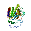
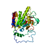
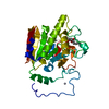
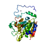
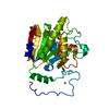
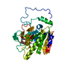
| Entry | Database: PDB / ID: 5cmf | ||||||
|---|---|---|---|---|---|---|---|
| Title | GTA mutant with mercury - E303A | ||||||
 Components Components | Histo-blood group ABO system transferase | ||||||
 Keywords Keywords | TRANSFERASE / Human ABO(H) blood group system / Glycosyltransferase / Double turn motif / Catalytic domain | ||||||
| Function / homology |  Function and homology information Function and homology informationfucosylgalactoside 3-alpha-galactosyltransferase / glycoprotein-fucosylgalactoside alpha-N-acetylgalactosaminyltransferase / glycoprotein-fucosylgalactoside alpha-N-acetylgalactosaminyltransferase activity / fucosylgalactoside 3-alpha-galactosyltransferase activity / ABO blood group biosynthesis / Golgi cisterna membrane / antigen binding / manganese ion binding / carbohydrate metabolic process / vesicle ...fucosylgalactoside 3-alpha-galactosyltransferase / glycoprotein-fucosylgalactoside alpha-N-acetylgalactosaminyltransferase / glycoprotein-fucosylgalactoside alpha-N-acetylgalactosaminyltransferase activity / fucosylgalactoside 3-alpha-galactosyltransferase activity / ABO blood group biosynthesis / Golgi cisterna membrane / antigen binding / manganese ion binding / carbohydrate metabolic process / vesicle / Golgi membrane / nucleotide binding / Golgi apparatus / extracellular region Similarity search - Function | ||||||
| Biological species |  Homo sapiens (human) Homo sapiens (human) | ||||||
| Method |  X-RAY DIFFRACTION / MOLECULAR REPLACEMENT / Resolution: 1.95 Å X-RAY DIFFRACTION / MOLECULAR REPLACEMENT / Resolution: 1.95 Å | ||||||
 Authors Authors | Gagnon, S.M.L. / Blackler, R.J. | ||||||
 Citation Citation | Journal: Glycobiology / Year: 2017 Title: Glycosyltransfer in mutants of putative catalytic residue Glu303 of the human ABO(H) A and B blood group glycosyltransferases GTA and GTB proceeds through a labile active site. Authors: Blackler, R.J. / Gagnon, S.M. / Polakowski, R. / Rose, N.L. / Zheng, R.B. / Letts, J.A. / Johal, A.R. / Schuman, B. / Borisova, S.N. / Palcic, M.M. / Evans, S.V. | ||||||
| History |
|
- Structure visualization
| Structure viewer | Molecule: MolmilJmol/JSmol |
|---|
- Downloads & links
-Download
| PDBx/mmCIF format | 5cmf.cif.gz | 72.4 KB | Display | PDBx/mmCIF format |
|---|---|---|---|---|
| PDB format | pdb5cmf.ent.gz | 51.1 KB | Display | PDB format |
| PDBx/mmJSON format | 5cmf.json.gz | Tree view | PDBx/mmJSON format | |
| Others |  Other downloads Other downloads |
-Validation report
| Arichive directory | https://data.pdbj.org/pub/pdb/validation_reports/cm/5cmfftp://data.pdbj.org/pub/pdb/validation_reports/cm/5cmf | HTTPS FTP |
|---|
-Related structure data
| Related structure data | 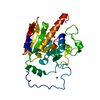 5cmgC 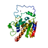 5cmhC 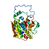 5cmiC 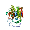 5cmjC 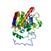 5cqlC 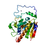 5cqmC 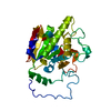 5cqnC 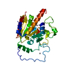 5cqoC 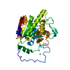 5cqpC 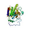 1lz0S C: citing same article ( S: Starting model for refinement |
|---|---|
| Similar structure data |


-Links
 PDBj
PDBj


- Assembly
| Deposited unit | 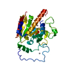
| |||||||||
|---|---|---|---|---|---|---|---|---|---|---|
| 1 |
| |||||||||
| Unit cell |
| |||||||||
| Components on special symmetry positions |
|
-Components
| #1: Protein | Mass: 33088.285 Da / Num. of mol.: 1 / Fragment: Catalytic domain (UNP residues 64-345) / Mutation: E303A Source method: isolated from a genetically manipulated source Source: (gene. exp.) Homo sapiens (human) / Gene: ABO / Production host:  References: UniProt: P16442, glycoprotein-fucosylgalactoside alpha-N-acetylgalactosaminyltransferase, fucosylgalactoside 3-alpha-galactosyltransferase | ||
|---|---|---|---|
| #2: Chemical | ChemComp-HG /   Mass: 200.590 Da / Num. of mol.: 4 / Source method: obtained synthetically / Formula: Hg Mass: 200.590 Da / Num. of mol.: 4 / Source method: obtained synthetically / Formula: Hg#3: Water | ChemComp-HOH / |  Mass: 18.015 Da / Num. of mol.: 118 / Source method: isolated from a natural source / Formula: H2O Mass: 18.015 Da / Num. of mol.: 118 / Source method: isolated from a natural source / Formula: H2O |
-Experimental details
-Experiment
| Experiment | Method: X-RAY DIFFRACTION / Number of used crystals: 1 |
|---|
- Sample preparation
| Crystal | Density Matthews: 2.33 Å3/Da / Density % sol: 47.2 % |
|---|---|
| Crystal grow | Temperature: 291 K / Method: vapor diffusion, hanging drop Details: 10 ul drops with 6-8 mg/ml protein, 70 mM N-(2-acetamido)-2-iminodiacetic acid (ADA) pH 7.5, 50 mM, sodium acetate pH 4.6, 40 mM NaCl, 5-8 mM MnCl2, 2.5% (v/v) 2-methyl-2,4-pentanediol (MPD) ...Details: 10 ul drops with 6-8 mg/ml protein, 70 mM N-(2-acetamido)-2-iminodiacetic acid (ADA) pH 7.5, 50 mM, sodium acetate pH 4.6, 40 mM NaCl, 5-8 mM MnCl2, 2.5% (v/v) 2-methyl-2,4-pentanediol (MPD), 5%(v/v) glycerol, 2%(w/v) PEG 4000, and 0.3-0.5 mM 3-chloromercuri-2-methoxypropylurea suspended over 1 ml of a resevoir solution: 50 mM ADA pH 7.5, 10 mM MnCl2, 100 mM ammonium sulfate, 5%(v/v) MPD, 10%(v/v) glycerol, and 8-10%(w/v) PEG 4000 |
-Data collection
| Diffraction | Mean temperature: 113 K | |||||||||||||||||||||||||||||||||||||||||||||||||||||||||||||||||||||||||||||||||||||||||||||||||||
|---|---|---|---|---|---|---|---|---|---|---|---|---|---|---|---|---|---|---|---|---|---|---|---|---|---|---|---|---|---|---|---|---|---|---|---|---|---|---|---|---|---|---|---|---|---|---|---|---|---|---|---|---|---|---|---|---|---|---|---|---|---|---|---|---|---|---|---|---|---|---|---|---|---|---|---|---|---|---|---|---|---|---|---|---|---|---|---|---|---|---|---|---|---|---|---|---|---|---|---|---|
| Diffraction source | Source: ROTATING ANODE / Type: RIGAKU / Wavelength: 1.5418 Å | |||||||||||||||||||||||||||||||||||||||||||||||||||||||||||||||||||||||||||||||||||||||||||||||||||
| Detector | Type: RIGAKU RAXIS IV++ / Detector: IMAGE PLATE / Date: Nov 1, 2011 | |||||||||||||||||||||||||||||||||||||||||||||||||||||||||||||||||||||||||||||||||||||||||||||||||||
| Radiation | Protocol: SINGLE WAVELENGTH / Monochromatic (M) / Laue (L): M / Scattering type: x-ray | |||||||||||||||||||||||||||||||||||||||||||||||||||||||||||||||||||||||||||||||||||||||||||||||||||
| Radiation wavelength | Wavelength: 1.5418 Å / Relative weight: 1 | |||||||||||||||||||||||||||||||||||||||||||||||||||||||||||||||||||||||||||||||||||||||||||||||||||
| Reflection | Resolution: 1.95→74.54 Å / Num. obs: 21791 / % possible obs: 94.9 % / Redundancy: 3.86 % / Rmerge(I) obs: 0.056 / Χ2: 0.83 / Net I/σ(I): 10.4 / Num. measured all: 84746 / Scaling rejects: 636 | |||||||||||||||||||||||||||||||||||||||||||||||||||||||||||||||||||||||||||||||||||||||||||||||||||
| Reflection shell | Diffraction-ID: 1
|
- Processing
| Software |
| |||||||||||||||||||||||||||||||||||||||||||||||||||||||||||||||||||||||||||
|---|---|---|---|---|---|---|---|---|---|---|---|---|---|---|---|---|---|---|---|---|---|---|---|---|---|---|---|---|---|---|---|---|---|---|---|---|---|---|---|---|---|---|---|---|---|---|---|---|---|---|---|---|---|---|---|---|---|---|---|---|---|---|---|---|---|---|---|---|---|---|---|---|---|---|---|---|
| Refinement | Method to determine structure: MOLECULAR REPLACEMENT Starting model: PDB entry 1LZ0 Resolution: 1.95→74.54 Å / Cor.coef. Fo:Fc: 0.958 / Cor.coef. Fo:Fc free: 0.939 / SU B: 4.967 / SU ML: 0.134 / Cross valid method: THROUGHOUT / σ(F): 0 / ESU R: 0.187 / ESU R Free: 0.168 / Stereochemistry target values: MAXIMUM LIKELIHOOD / Details: HYDROGENS HAVE BEEN ADDED IN THE RIDING POSITIONS
| |||||||||||||||||||||||||||||||||||||||||||||||||||||||||||||||||||||||||||
| Solvent computation | Ion probe radii: 0.8 Å / Shrinkage radii: 0.8 Å / VDW probe radii: 1.2 Å / Solvent model: MASK | |||||||||||||||||||||||||||||||||||||||||||||||||||||||||||||||||||||||||||
| Displacement parameters | Biso max: 86.3 Å2 / Biso mean: 40.613 Å2 / Biso min: 25.44 Å2
| |||||||||||||||||||||||||||||||||||||||||||||||||||||||||||||||||||||||||||
| Refinement step | Cycle: final / Resolution: 1.95→74.54 Å
| |||||||||||||||||||||||||||||||||||||||||||||||||||||||||||||||||||||||||||
| Refine LS restraints |
| |||||||||||||||||||||||||||||||||||||||||||||||||||||||||||||||||||||||||||
| LS refinement shell | Resolution: 1.95→2.001 Å / Total num. of bins used: 20
|
