 Movie
Movie Controller


+ Open data

- Basic information
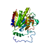
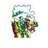
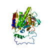
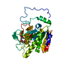
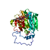
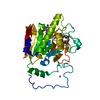
| Entry | Database: PDB / ID: 1lzi | |||||||||
|---|---|---|---|---|---|---|---|---|---|---|
| Title | Glycosyltransferase A + UDP + H antigen acceptor | |||||||||
 Components Components | Glycosyltransferase A | |||||||||
 Keywords Keywords | TRANSFERASE / GLYCOPROTEIN / TRANSMEMBRANE / SIGNAL-ANCHOR / BLOOD GROUP ANTIGEN | |||||||||
| Function / homology |  Function and homology information Function and homology informationfucosylgalactoside 3-alpha-galactosyltransferase / glycoprotein-fucosylgalactoside alpha-N-acetylgalactosaminyltransferase / glycoprotein-fucosylgalactoside alpha-N-acetylgalactosaminyltransferase activity / fucosylgalactoside 3-alpha-galactosyltransferase activity / ABO blood group biosynthesis / Golgi cisterna membrane / antigen binding / manganese ion binding / carbohydrate metabolic process / vesicle ...fucosylgalactoside 3-alpha-galactosyltransferase / glycoprotein-fucosylgalactoside alpha-N-acetylgalactosaminyltransferase / glycoprotein-fucosylgalactoside alpha-N-acetylgalactosaminyltransferase activity / fucosylgalactoside 3-alpha-galactosyltransferase activity / ABO blood group biosynthesis / Golgi cisterna membrane / antigen binding / manganese ion binding / carbohydrate metabolic process / vesicle / Golgi membrane / nucleotide binding / Golgi apparatus / extracellular region Similarity search - Function | |||||||||
| Biological species |  Homo sapiens (human) Homo sapiens (human) | |||||||||
| Method |  X-RAY DIFFRACTION / SYNCHROTRON / MOLECULAR REPLACEMENT / Resolution: 1.35 Å X-RAY DIFFRACTION / SYNCHROTRON / MOLECULAR REPLACEMENT / Resolution: 1.35 Å | |||||||||
 Authors Authors | Patenaude, S.I. / Seto, N.O.L. / Borisova, S.N. / Szpacenko, A. / Marcus, S.L. / Palcic, M.M. / Evans, S.V. | |||||||||
 Citation Citation | Journal: Nat.Struct.Biol. / Year: 2002 Title: The structural basis for specificity in human ABO(H) blood group biosynthesis. Authors: Patenaude, S.I. / Seto, N.O. / Borisova, S.N. / Szpacenko, A. / Marcus, S.L. / Palcic, M.M. / Evans, S.V. | |||||||||
| History |
|
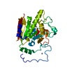
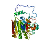
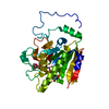
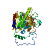
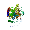
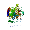
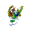
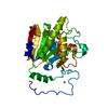
- Structure visualization
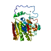
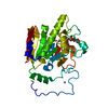
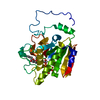
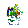
| Structure viewer | Molecule: MolmilJmol/JSmol |
|---|
- Downloads & links
-Download
| PDBx/mmCIF format | 1lzi.cif.gz | 82.5 KB | Display | PDBx/mmCIF format |
|---|---|---|---|---|
| PDB format | pdb1lzi.ent.gz | 59.5 KB | Display | PDB format |
| PDBx/mmJSON format | 1lzi.json.gz | Tree view | PDBx/mmJSON format | |
| Others |  Other downloads Other downloads |
-Validation report
| Arichive directory | https://data.pdbj.org/pub/pdb/validation_reports/lz/1lziftp://data.pdbj.org/pub/pdb/validation_reports/lz/1lzi | HTTPS FTP |
|---|
-Related structure data


-Links
 PDBj
PDBj


- Assembly
| Deposited unit | 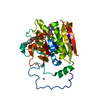
| ||||||||
|---|---|---|---|---|---|---|---|---|---|
| 1 |
| ||||||||
| 2 | 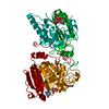
| ||||||||
| Unit cell |
|
-Components
-Protein / Sugars , 2 types, 2 molecules A
| #1: Protein | Mass: 34047.324 Da / Num. of mol.: 1 / Fragment: Catalytic domain, (RESIDUES 64-354) Source method: isolated from a genetically manipulated source Details: N-terminal truncation / Source: (gene. exp.) Homo sapiens (human) / Production host:  References: UniProt: P16442, glycoprotein-fucosylgalactoside alpha-N-acetylgalactosaminyltransferase |
|---|---|
| #2: Polysaccharide | alpha-L-fucopyranose-(1-2)-hexyl beta-D-galactopyranoside Type: oligosaccharide / Mass: 410.456 Da / Num. of mol.: 1 Source method: isolated from a genetically manipulated source |
-Non-polymers , 4 types, 401 molecules 


| #3: Chemical | ChemComp-HG /  Mass: 200.590 Da / Num. of mol.: 5 / Source method: obtained synthetically / Formula: Hg Mass: 200.590 Da / Num. of mol.: 5 / Source method: obtained synthetically / Formula: Hg#4: Chemical | ChemComp-MN / |  Mass: 54.938 Da / Num. of mol.: 1 / Source method: obtained synthetically / Formula: Mn Mass: 54.938 Da / Num. of mol.: 1 / Source method: obtained synthetically / Formula: Mn#5: Chemical | ChemComp-UDP / |  Type: RNA linking / Mass: 404.161 Da / Num. of mol.: 1 / Source method: obtained synthetically / Formula: C9H14N2O12P2 / Comment: UDP*YM Type: RNA linking / Mass: 404.161 Da / Num. of mol.: 1 / Source method: obtained synthetically / Formula: C9H14N2O12P2 / Comment: UDP*YM#6: Water | ChemComp-HOH / | Mass: 18.015 Da / Num. of mol.: 394 / Source method: isolated from a natural source / Formula: H2O |
|---|
-Experimental details
-Experiment
| Experiment | Method: X-RAY DIFFRACTION / Number of used crystals: 1 |
|---|
- Sample preparation
| Crystal | Density Matthews: 2.32 Å3/Da / Density % sol: 46.94 % | ||||||||||||||||||||||||||||||||||||||||||||||||||||||||||||||||||||||||||||||||||||||||||||||||||||||||||||||||
|---|---|---|---|---|---|---|---|---|---|---|---|---|---|---|---|---|---|---|---|---|---|---|---|---|---|---|---|---|---|---|---|---|---|---|---|---|---|---|---|---|---|---|---|---|---|---|---|---|---|---|---|---|---|---|---|---|---|---|---|---|---|---|---|---|---|---|---|---|---|---|---|---|---|---|---|---|---|---|---|---|---|---|---|---|---|---|---|---|---|---|---|---|---|---|---|---|---|---|---|---|---|---|---|---|---|---|---|---|---|---|---|---|---|
| Crystal grow | Temperature: 291 K / Method: vapor diffusion, hanging drop / pH: 7.5 Details: ADA, manganese chloride, ammonium sulfate, MPD, glycerol, PEG 4000, pH 7.5, VAPOR DIFFUSION, HANGING DROP, temperature 291K | ||||||||||||||||||||||||||||||||||||||||||||||||||||||||||||||||||||||||||||||||||||||||||||||||||||||||||||||||
| Crystal grow | *PLUS Temperature: 18 ℃ | ||||||||||||||||||||||||||||||||||||||||||||||||||||||||||||||||||||||||||||||||||||||||||||||||||||||||||||||||
| Components of the solutions | *PLUS
|
-Data collection
| Diffraction | Mean temperature: 100 K |
|---|---|
| Diffraction source | Source: SYNCHROTRON / Site: NSLS  / Beamline: X8C / Wavelength: 1.15 Å / Beamline: X8C / Wavelength: 1.15 Å |
| Detector | Type: ADSC QUANTUM 4 / Detector: CCD |
| Radiation | Protocol: SINGLE WAVELENGTH / Monochromatic (M) / Laue (L): M / Scattering type: x-ray |
| Radiation wavelength | Wavelength: 1.15 Å / Relative weight: 1 |
| Reflection | Resolution: 1.35→50 Å / Num. all: 66876 / Num. obs: 66876 / Observed criterion σ(F): 0 / Observed criterion σ(I): 0 / Limit h max: 38 / Limit h min: 0 / Limit k max: 100 / Limit k min: 0 / Limit l max: 59 / Limit l min: 0 / Observed criterion F max: 438683.59 / Observed criterion F min: 0.32 |
| Reflection shell | Resolution: 1.35→1.41 Å / % possible all: 69.5 |
| Reflection | *PLUS Lowest resolution: 50 Å / % possible obs: 95.5 % / Rmerge(I) obs: 0.05 |
| Reflection shell | *PLUS % possible obs: 69.5 % / Rmerge(I) obs: 0.224 / Mean I/σ(I) obs: 4.99 |
- Processing
| Software |
| ||||||||||||||||||||||||||||||||||||||||||||||||||||||||||||||||||||||||||||||||||||||||||
|---|---|---|---|---|---|---|---|---|---|---|---|---|---|---|---|---|---|---|---|---|---|---|---|---|---|---|---|---|---|---|---|---|---|---|---|---|---|---|---|---|---|---|---|---|---|---|---|---|---|---|---|---|---|---|---|---|---|---|---|---|---|---|---|---|---|---|---|---|---|---|---|---|---|---|---|---|---|---|---|---|---|---|---|---|---|---|---|---|---|---|---|
| Refinement | Method to determine structure: MOLECULAR REPLACEMENT / Resolution: 1.35→20 Å / Rfactor Rfree error: 0.003 / Occupancy max: 1 / Occupancy min: 0.5 / σ(F): 2 / Stereochemistry target values: Engh & Huber
| ||||||||||||||||||||||||||||||||||||||||||||||||||||||||||||||||||||||||||||||||||||||||||
| Solvent computation | Solvent model: CNS bulk solvent model used / Bsol: 77.2011 Å2 / ksol: 0.380403 e/Å3 | ||||||||||||||||||||||||||||||||||||||||||||||||||||||||||||||||||||||||||||||||||||||||||
| Displacement parameters | Biso max: 61.95 Å2 / Biso min: 7.62 Å2 | ||||||||||||||||||||||||||||||||||||||||||||||||||||||||||||||||||||||||||||||||||||||||||
| Refine analyze |
| ||||||||||||||||||||||||||||||||||||||||||||||||||||||||||||||||||||||||||||||||||||||||||
| Refinement step | Cycle: LAST / Resolution: 1.35→20 Å
| ||||||||||||||||||||||||||||||||||||||||||||||||||||||||||||||||||||||||||||||||||||||||||
| Refine LS restraints |
| ||||||||||||||||||||||||||||||||||||||||||||||||||||||||||||||||||||||||||||||||||||||||||
| LS refinement shell | Refine-ID: X-RAY DIFFRACTION
| ||||||||||||||||||||||||||||||||||||||||||||||||||||||||||||||||||||||||||||||||||||||||||
| Software | *PLUS Name: CNS / Classification: refinement | ||||||||||||||||||||||||||||||||||||||||||||||||||||||||||||||||||||||||||||||||||||||||||
| Refinement | *PLUS Lowest resolution: 20 Å / % reflection Rfree: 10 % / Rfactor all: 0.184 / Rfactor obs: 0.182 / Rfactor Rfree: 0.2 / Rfactor Rwork: 0.179 | ||||||||||||||||||||||||||||||||||||||||||||||||||||||||||||||||||||||||||||||||||||||||||
| Solvent computation | *PLUS | ||||||||||||||||||||||||||||||||||||||||||||||||||||||||||||||||||||||||||||||||||||||||||
| Displacement parameters | *PLUS | ||||||||||||||||||||||||||||||||||||||||||||||||||||||||||||||||||||||||||||||||||||||||||
| Refine LS restraints | *PLUS
|
