 Movie
Movie Controller


+ Open data

- Basic information
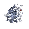
| Entry | Database: PDB / ID: 1hm7 | ||||||
|---|---|---|---|---|---|---|---|
| Title | N219L PENTALENENE SYNTHASE | ||||||
 Components Components | PENTALENENE SYNTHASE | ||||||
 Keywords Keywords | LYASE / sesquiterpene synthase / pentalenene / terpene / antibiotic biosynthesis | ||||||
| Function / homology |  Function and homology information Function and homology informationpentalenene synthase / pentalenene synthase activity / antibiotic biosynthetic process / metal ion binding Similarity search - Function | ||||||
| Biological species |  Streptomyces sp. (bacteria) Streptomyces sp. (bacteria) | ||||||
| Method |  X-RAY DIFFRACTION / FOURIER SYNTHESIS / Resolution: 2.9 Å X-RAY DIFFRACTION / FOURIER SYNTHESIS / Resolution: 2.9 Å | ||||||
 Authors Authors | Seemann, M. / Paschall, C.M. / Christianson, D.W. / Cane, D.E. | ||||||
 Citation Citation | Journal: J.Am.Chem.Soc. / Year: 2002 Title: Pentalenene synthase. Analysis of active site residues by site-directed mutagenesis. Authors: Seemann, M. / Zhai, G. / de Kraker, J.W. / Paschall, C.M. / Christianson, D.W. / Cane, D.E. | ||||||
| History |
|
- Structure visualization
| Structure viewer | Molecule: MolmilJmol/JSmol |
|---|
- Downloads & links
-Download
| PDBx/mmCIF format | 1hm7.cif.gz | 125.9 KB | Display | PDBx/mmCIF format |
|---|---|---|---|---|
| PDB format | pdb1hm7.ent.gz | 99 KB | Display | PDB format |
| PDBx/mmJSON format | 1hm7.json.gz | Tree view | PDBx/mmJSON format | |
| Others |  Other downloads Other downloads |
-Validation report
| Arichive directory | https://data.pdbj.org/pub/pdb/validation_reports/hm/1hm7ftp://data.pdbj.org/pub/pdb/validation_reports/hm/1hm7 | HTTPS FTP |
|---|
-Related structure data
| Related structure data |  1hm4SC S: Starting model for refinement C: citing same article ( |
|---|---|
| Similar structure data |


-Links
 PDBj
PDBj
- Assembly
| Deposited unit | 
| ||||||||||
|---|---|---|---|---|---|---|---|---|---|---|---|
| 1 | 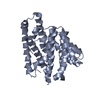
| ||||||||||
| 2 | 
| ||||||||||
| Unit cell |
| ||||||||||
| Details | The biological assembly is a monomer |
-Components
| #1: Protein | Mass: 37918.273 Da / Num. of mol.: 2 / Mutation: N219L Source method: isolated from a genetically manipulated source Source: (gene. exp.) Streptomyces sp. (bacteria) / Strain: UC5319 / Production host: #2: Water | ChemComp-HOH / |  Mass: 18.015 Da / Num. of mol.: 26 / Source method: isolated from a natural source / Formula: H2O Mass: 18.015 Da / Num. of mol.: 26 / Source method: isolated from a natural source / Formula: H2OHas protein modification | Y | |
|---|
-Experimental details
-Experiment
| Experiment | Method: X-RAY DIFFRACTION / Number of used crystals: 1 |
|---|
- Sample preparation
| Crystal | Density Matthews: 3.4 Å3/Da / Density % sol: 64 % | ||||||||||||||||||||||||||||||
|---|---|---|---|---|---|---|---|---|---|---|---|---|---|---|---|---|---|---|---|---|---|---|---|---|---|---|---|---|---|---|---|
| Crystal grow | Temperature: 298 K / Method: vapor diffusion, sitting drop / pH: 7 Details: (NH4)2SO4, ethanol, MgCl2, Hepes pH 7.0, farnesyl diphosphate, VAPOR DIFFUSION, SITTING DROP at 298K | ||||||||||||||||||||||||||||||
| Crystal grow | *PLUS | ||||||||||||||||||||||||||||||
| Components of the solutions | *PLUS
|
-Data collection
| Diffraction | Mean temperature: 200 K |
|---|---|
| Diffraction source | Source: ROTATING ANODE / Type: RIGAKU RU200 / Wavelength: 1.5418 Å |
| Detector | Type: RIGAKU RAXIS IIC / Detector: IMAGE PLATE / Date: Apr 4, 2000 / Details: mirrors |
| Radiation | Monochromator: Yale mirrors / Protocol: SINGLE WAVELENGTH / Monochromatic (M) / Laue (L): M / Scattering type: x-ray |
| Radiation wavelength | Wavelength: 1.5418 Å / Relative weight: 1 |
| Reflection | Resolution: 2.9→20 Å / Num. all: 23110 / Num. obs: 23110 / % possible obs: 96.8 % / Observed criterion σ(F): 0 / Observed criterion σ(I): 0 / Redundancy: 2.76 % / Biso Wilson estimate: 45.22 Å2 / Rmerge(I) obs: 0.083 / Rsym value: 0.083 / Net I/σ(I): 11.87 |
| Reflection shell | Resolution: 2.9→3 Å / Redundancy: 1 % / Rmerge(I) obs: 0.404 / Mean I/σ(I) obs: 2.5 / Num. unique all: 2022 / Rsym value: 0.404 / % possible all: 95.8 |
| Reflection | *PLUS Highest resolution: 2.9 Å / % possible obs: 97 % / Num. measured all: 63768 |
- Processing
| Software |
| |||||||||||||||||||||||||
|---|---|---|---|---|---|---|---|---|---|---|---|---|---|---|---|---|---|---|---|---|---|---|---|---|---|---|
| Refinement | Method to determine structure: FOURIER SYNTHESIS Starting model: PDB ENTRY 1HM4 Resolution: 2.9→20 Å / Isotropic thermal model: ANISOTROPIC / Cross valid method: THROUGHOUT / σ(F): 0 / σ(I): 0 / Stereochemistry target values: ENGH & HUBER
| |||||||||||||||||||||||||
| Displacement parameters |
| |||||||||||||||||||||||||
| Refine analyze |
| |||||||||||||||||||||||||
| Refinement step | Cycle: LAST / Resolution: 2.9→20 Å
| |||||||||||||||||||||||||
| Refine LS restraints |
| |||||||||||||||||||||||||
| LS refinement shell | Resolution: 2.9→3 Å
| |||||||||||||||||||||||||
| Refinement | *PLUS Lowest resolution: 20 Å / Rfactor Rwork: 0.263 | |||||||||||||||||||||||||
| Solvent computation | *PLUS | |||||||||||||||||||||||||
| Displacement parameters | *PLUS | |||||||||||||||||||||||||
| Refine LS restraints | *PLUS
|
