 Movie
Movie Controller


+ Open data

- Basic information
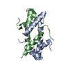
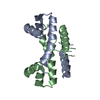
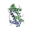
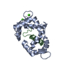
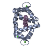
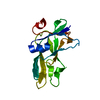
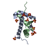
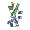
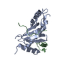
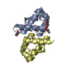
| Entry | Database: PDB / ID: 1ety | ||||||
|---|---|---|---|---|---|---|---|
| Title | THE CRYSTAL STRUCTURE OF E. COLI WILD-TYPE FIS | ||||||
 Components Components | FACTOR FOR INVERSION STIMULATION | ||||||
 Keywords Keywords | TRANSCRIPTION ACTIVATOR / DNA-BINDING PROTEIN | ||||||
| Function / homology |  Function and homology information Function and homology informationinvertasome / positive regulation of DNA recombination / sequence-specific DNA binding, bending / provirus excision / nucleoid / DNA-binding transcription repressor activity / DNA-binding transcription activator activity / chromosome organization / core promoter sequence-specific DNA binding / protein-DNA complex ...invertasome / positive regulation of DNA recombination / sequence-specific DNA binding, bending / provirus excision / nucleoid / DNA-binding transcription repressor activity / DNA-binding transcription activator activity / chromosome organization / core promoter sequence-specific DNA binding / protein-DNA complex / response to radiation / nucleosome / sequence-specific DNA binding / transcription cis-regulatory region binding / regulation of DNA-templated transcription / DNA-templated transcription / protein homodimerization activity / DNA binding / cytosol Similarity search - Function | ||||||
| Biological species |  | ||||||
| Method |  X-RAY DIFFRACTION / Resolution: 2 Å X-RAY DIFFRACTION / Resolution: 2 Å | ||||||
 Authors Authors | Cheng, Y.S. / Yang, W.Z. / Johnson, R.C. / Yuan, H.S. | ||||||
 Citation Citation | Journal: J.Mol.Biol. / Year: 2000 Title: Structural analysis of the transcriptional activation on Fis: crystal structures of six Fis mutants with different activation properties. Authors: Cheng, Y.S. / Yang, W.Z. / Johnson, R.C. / Yuan, H.S. | ||||||
| History |
| ||||||
| Remark 300 | THIS ENTRY CONTAINS THE CRYSTALLOGRAPHIC ASYMMETRIC UNIT WHICH CONSISTS OF 2 CHAINS THAT GIVE ONE ... THIS ENTRY CONTAINS THE CRYSTALLOGRAPHIC ASYMMETRIC UNIT WHICH CONSISTS OF 2 CHAINS THAT GIVE ONE BIOLOGICAL DIMER MOLECULE. |
- Structure visualization
| Structure viewer | Molecule: MolmilJmol/JSmol |
|---|
- Downloads & links
-Download
| PDBx/mmCIF format | 1ety.cif.gz | 43.6 KB | Display | PDBx/mmCIF format |
|---|---|---|---|---|
| PDB format | pdb1ety.ent.gz | 31.5 KB | Display | PDB format |
| PDBx/mmJSON format | 1ety.json.gz | Tree view | PDBx/mmJSON format | |
| Others |  Other downloads Other downloads |
-Validation report
| Arichive directory | https://data.pdbj.org/pub/pdb/validation_reports/et/1etyftp://data.pdbj.org/pub/pdb/validation_reports/et/1ety | HTTPS FTP |
|---|
-Related structure data
| Related structure data |  1etkC 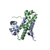 1etoC  1etqC  1etvC  1etwC  1etxC C: citing same article ( |
|---|---|
| Similar structure data |


-Links
 PDBj
PDBj- Assembly
| Deposited unit | 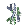
| ||||||||
|---|---|---|---|---|---|---|---|---|---|
| 1 |
| ||||||||
| Unit cell |
| ||||||||
| Details | The biological assembly is a dimer constructed from chain A and a symmetry partner (chain B) generated by the non-crystallographic two-fold |
-Components
| #1: Protein | Mass: 11252.918 Da / Num. of mol.: 2 Source method: isolated from a genetically manipulated source Source: (gene. exp.) #2: Water | ChemComp-HOH / |  Mass: 18.015 Da / Num. of mol.: 111 / Source method: isolated from a natural source / Formula: H2O Mass: 18.015 Da / Num. of mol.: 111 / Source method: isolated from a natural source / Formula: H2O |
|---|
-Experimental details
-Experiment
| Experiment | Method: X-RAY DIFFRACTION / Number of used crystals: 1 |
|---|
- Sample preparation
| Crystal | Density Matthews: 2.07 Å3/Da / Density % sol: 40.49 % | ||||||||||||||||||||||||
|---|---|---|---|---|---|---|---|---|---|---|---|---|---|---|---|---|---|---|---|---|---|---|---|---|---|
| Crystal grow | Temperature: 298 K / Method: vapor diffusion, hanging drop / pH: 6.5 Details: 10 mg/ml protein, 0.5 M sodium chloride, 0.1 M sodium acetate, 0.05 M sodium cacodylate(pH 6.5), 15% PEG8000, VAPOR DIFFUSION, HANGING DROP, temperature 298K | ||||||||||||||||||||||||
| Crystal grow | *PLUS | ||||||||||||||||||||||||
| Components of the solutions | *PLUS
|
-Data collection
| Diffraction | Mean temperature: 120 K |
|---|---|
| Diffraction source | Source: ROTATING ANODE / Type: RIGAKU RU300 / Wavelength: 1.5418 |
| Detector | Type: RIGAKU RAXIS II / Detector: IMAGE PLATE / Date: Sep 2, 1999 / Details: mirrors |
| Radiation | Protocol: SINGLE WAVELENGTH / Monochromatic (M) / Laue (L): M / Scattering type: x-ray |
| Radiation wavelength | Wavelength: 1.5418 Å / Relative weight: 1 |
| Reflection | Resolution: 2→40 Å / Num. all: 70772 / Num. obs: 13174 / % possible obs: 99.7 % / Observed criterion σ(F): 0 / Observed criterion σ(I): 0 / Redundancy: 5.37 % / Biso Wilson estimate: 17.7 Å2 / Rsym value: 0.062 / Net I/σ(I): 25.1 |
| Reflection shell | Resolution: 2→2.07 Å / Redundancy: 6.39 % / Num. unique all: 1279 / Rsym value: 0.254 / % possible all: 99.6 |
| Reflection | *PLUS Num. measured all: 70772 / Rmerge(I) obs: 0.062 |
| Reflection shell | *PLUS % possible obs: 99.6 % / Rmerge(I) obs: 0.254 / Mean I/σ(I) obs: 6.4 |
- Processing
| Software |
| ||||||||||||||||||||||||||||||||||||
|---|---|---|---|---|---|---|---|---|---|---|---|---|---|---|---|---|---|---|---|---|---|---|---|---|---|---|---|---|---|---|---|---|---|---|---|---|---|
| Refinement | Starting model: The wild-type FIS Resolution: 2→19.4 Å / Rfactor Rfree error: 0.008 / Data cutoff high rms absF: 450184.2 / Isotropic thermal model: restrained / σ(F): 0 / σ(I): 0 / Stereochemistry target values: Engh & Huber
| ||||||||||||||||||||||||||||||||||||
| Solvent computation | Solvent model: flat model / Bsol: 50.0204 Å2 / ksol: 0.342513 e/Å3 | ||||||||||||||||||||||||||||||||||||
| Displacement parameters | Biso mean: 26.8 Å2
| ||||||||||||||||||||||||||||||||||||
| Refine analyze |
| ||||||||||||||||||||||||||||||||||||
| Refinement step | Cycle: LAST / Resolution: 2→19.4 Å
| ||||||||||||||||||||||||||||||||||||
| Refine LS restraints |
| ||||||||||||||||||||||||||||||||||||
| LS refinement shell | Resolution: 2→2.13 Å / Rfactor Rfree error: 0.021 / Total num. of bins used: 6
| ||||||||||||||||||||||||||||||||||||
| Xplor file |
| ||||||||||||||||||||||||||||||||||||
| Software | *PLUS Name: CNS / Version: 0.9 / Classification: refinement | ||||||||||||||||||||||||||||||||||||
| Refine LS restraints | *PLUS
|
