 Movie
Movie Controller

[English] 日本語
 Yorodumi
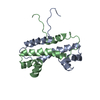
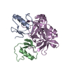
- PDB-1f36: THE CRYSTAL STRUCTURE OF FIS MUTANT K36E REVEALS THAT THE TRANSAC... -
+ Open data

- Basic information
| Entry | Database: PDB / ID: 1f36 | ||||||
|---|---|---|---|---|---|---|---|
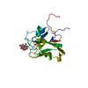
| Title | THE CRYSTAL STRUCTURE OF FIS MUTANT K36E REVEALS THAT THE TRANSACTIVATION REGION OF THE FIS PROTEIN CONTAINS EXTENDED MOBILE BETA-HAIRPIN ARMS | ||||||
 Components Components | FIS | ||||||
 Keywords Keywords | TRANSACTIVATION REGION / DNA-BINDING PROTEIN / PROTEIN-PROTEIN INTERACTION DOMAIN | ||||||
| Function / homology |  Function and homology information Function and homology informationinvertasome / positive regulation of DNA recombination / sequence-specific DNA binding, bending / provirus excision / nucleoid / DNA-binding transcription repressor activity / DNA-binding transcription activator activity / chromosome organization / core promoter sequence-specific DNA binding / protein-DNA complex ...invertasome / positive regulation of DNA recombination / sequence-specific DNA binding, bending / provirus excision / nucleoid / DNA-binding transcription repressor activity / DNA-binding transcription activator activity / chromosome organization / core promoter sequence-specific DNA binding / protein-DNA complex / response to radiation / nucleosome / sequence-specific DNA binding / transcription cis-regulatory region binding / regulation of DNA-templated transcription / DNA-templated transcription / protein homodimerization activity / DNA binding / cytosol Similarity search - Function | ||||||
| Biological species |  | ||||||
| Method |  X-RAY DIFFRACTION / MOLECULAR REPLACEMENT / Resolution: 2.65 Å X-RAY DIFFRACTION / MOLECULAR REPLACEMENT / Resolution: 2.65 Å | ||||||
 Authors Authors | Safo, M.K. / Yuan, H.S. | ||||||
 Citation Citation | Journal: EMBO J. / Year: 1997 Title: The transactivation region of the fis protein that controls site-specific DNA inversion contains extended mobile beta-hairpin arms. Authors: Safo, M.K. / Yang, W.Z. / Corselli, L. / Cramton, S.E. / Yuan, H.S. / Johnson, R.C. | ||||||
| History |
|
- Structure visualization
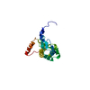
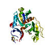
| Structure viewer | Molecule: MolmilJmol/JSmol |
|---|
- Downloads & links
-Download
| PDBx/mmCIF format | 1f36.cif.gz | 54 KB | Display | PDBx/mmCIF format |
|---|---|---|---|---|
| PDB format | pdb1f36.ent.gz | 40.7 KB | Display | PDB format |
| PDBx/mmJSON format | 1f36.json.gz | Tree view | PDBx/mmJSON format | |
| Others |  Other downloads Other downloads |
-Validation report
| Arichive directory | https://data.pdbj.org/pub/pdb/validation_reports/f3/1f36ftp://data.pdbj.org/pub/pdb/validation_reports/f3/1f36 | HTTPS FTP |
|---|
-Related structure data
| Similar structure data |
|---|


-Links
 PDBj
PDBj- Assembly
| Deposited unit | 
| |||||||||
|---|---|---|---|---|---|---|---|---|---|---|
| 1 |
| |||||||||
| Unit cell |
| |||||||||
| Noncrystallographic symmetry (NCS) | NCS domain:
|
-Components
| #1: Protein | Mass: 11252.853 Da / Num. of mol.: 2 / Mutation: K36E Source method: isolated from a genetically manipulated source Source: (gene. exp.) #2: Water | ChemComp-HOH / |  Mass: 18.015 Da / Num. of mol.: 22 / Source method: isolated from a natural source / Formula: H2O Mass: 18.015 Da / Num. of mol.: 22 / Source method: isolated from a natural source / Formula: H2O |
|---|
-Experimental details
-Experiment
| Experiment | Method: X-RAY DIFFRACTION / Number of used crystals: 1 |
|---|
- Sample preparation
| Crystal | Density Matthews: 2.56 Å3/Da / Density % sol: 52 % Description: SEARCH MODEL INCLUDED RESIDUES 24 - 98 OF THE DIMER AND THE CORRESPONDING TETRAPEPTIDE CHAINS (RESIDUES 10 - 13). | ||||||||||||||||||||
|---|---|---|---|---|---|---|---|---|---|---|---|---|---|---|---|---|---|---|---|---|---|
| Crystal grow | Method: vapor diffusion, hanging drop / pH: 8.2 Details: CRYSTALS GROWN BY HANGING DROP VAPOR DIFFUSION FROM A SOLUTION OF 20 MG/ML PROTEIN, 500 MM NACL, 10 MM TRIS-HCL, PH 8.2, 2 M NAFORMATE, AGAINST 4 M NAFORMATE AS THE RESERVOIR, vapor diffusion - hanging drop | ||||||||||||||||||||
| Crystal grow | *PLUS Method: vapor diffusion, hanging drop | ||||||||||||||||||||
| Components of the solutions | *PLUS
|
-Data collection
| Diffraction | Mean temperature: 298 K |
|---|---|
| Diffraction source | Source: ROTATING ANODE / Type: RIGAKU RUH3R / Wavelength: 1.5418 |
| Detector | Type: RIGAKU RAXIS II / Detector: IMAGE PLATE / Date: Jul 16, 1996 / Details: Y |
| Radiation | Monochromatic (M) / Laue (L): M / Scattering type: x-ray |
| Radiation wavelength | Wavelength: 1.5418 Å / Relative weight: 1 |
| Reflection | Resolution: 2.65→50 Å / Num. obs: 7139 / % possible obs: 93 % / Observed criterion σ(I): 0 / Redundancy: 11.1 % / Biso Wilson estimate: 54.6 Å2 / Rmerge(I) obs: 0.069 / Rsym value: 0.069 |
| Reflection shell | Resolution: 2.65→2.75 Å / Redundancy: 2 % / Rmerge(I) obs: 0.301 / Mean I/σ(I) obs: 1.63 / % possible all: 55.6 |
| Reflection | *PLUS Num. measured all: 79532 |
| Reflection shell | *PLUS % possible obs: 55.6 % |
- Processing
| Software |
| ||||||||||||||||||||||||||||||||||||||||||||||||||||||||||||
|---|---|---|---|---|---|---|---|---|---|---|---|---|---|---|---|---|---|---|---|---|---|---|---|---|---|---|---|---|---|---|---|---|---|---|---|---|---|---|---|---|---|---|---|---|---|---|---|---|---|---|---|---|---|---|---|---|---|---|---|---|---|
| Refinement | Method to determine structure: MOLECULAR REPLACEMENT Starting model: ORTHORHOMBIC CRYSTAL FORM OF THE LYS36-GLU FIS STRUCTURE Resolution: 2.65→8 Å / σ(F): 1 Details: MET A 1 - ASP A 9 ARE DISORDERED IN THE PROTEIN STRUCTURE. MET B 1 - ASP B 9 ARE DISORDERED IN THE PROTEIN STRUCTURE. PREVIOUSLY DISORDERED REGION (RESIDUES 1 - 24) IN ALL THE KNOWN CRYSTAL ...Details: MET A 1 - ASP A 9 ARE DISORDERED IN THE PROTEIN STRUCTURE. MET B 1 - ASP B 9 ARE DISORDERED IN THE PROTEIN STRUCTURE. PREVIOUSLY DISORDERED REGION (RESIDUES 1 - 24) IN ALL THE KNOWN CRYSTAL STRUCTERS OF THE FIS PROTEIN IS NOW PARTLY RESOLVED IN THIS LYS36-GLU FIS MUTANT (RESIDUES 10 - 24) AS BETA HAIRPIN STRUCTURE.
| ||||||||||||||||||||||||||||||||||||||||||||||||||||||||||||
| Displacement parameters | Biso mean: 42.1 Å2 | ||||||||||||||||||||||||||||||||||||||||||||||||||||||||||||
| Refine analyze | Luzzati coordinate error obs: 0.3 Å / Luzzati d res low obs: 8 Å / Luzzati sigma a obs: 0.52 Å | ||||||||||||||||||||||||||||||||||||||||||||||||||||||||||||
| Refinement step | Cycle: LAST / Resolution: 2.65→8 Å
| ||||||||||||||||||||||||||||||||||||||||||||||||||||||||||||
| Refine LS restraints |
| ||||||||||||||||||||||||||||||||||||||||||||||||||||||||||||
| Refine LS restraints NCS |
| ||||||||||||||||||||||||||||||||||||||||||||||||||||||||||||
| LS refinement shell | Resolution: 2.65→2.77 Å / Total num. of bins used: 8
| ||||||||||||||||||||||||||||||||||||||||||||||||||||||||||||
| Xplor file |
| ||||||||||||||||||||||||||||||||||||||||||||||||||||||||||||
| Software | *PLUS Name: X-PLOR / Version: 3.1 / Classification: refinement | ||||||||||||||||||||||||||||||||||||||||||||||||||||||||||||
| Refinement | *PLUS | ||||||||||||||||||||||||||||||||||||||||||||||||||||||||||||
| Solvent computation | *PLUS | ||||||||||||||||||||||||||||||||||||||||||||||||||||||||||||
| Displacement parameters | *PLUS | ||||||||||||||||||||||||||||||||||||||||||||||||||||||||||||
| Refine LS restraints | *PLUS
| ||||||||||||||||||||||||||||||||||||||||||||||||||||||||||||
| LS refinement shell | *PLUS Rfactor obs: 0.312 |
