 Movie
Movie Controller


+ Open data

- Basic information
| Entry | Database: PDB / ID: 1ejb | ||||||
|---|---|---|---|---|---|---|---|
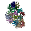
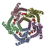
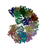
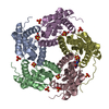
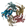
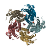
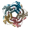
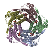
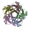
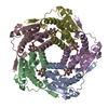
| Title | LUMAZINE SYNTHASE FROM SACCHAROMYCES CEREVISIAE | ||||||
 Components Components | LUMAZINE SYNTHASE | ||||||
 Keywords Keywords | TRANSFERASE / Lumazine synthase / Saccharomyces cerevisiae / X-ray structure analysis / Inhibitor complex / Vitamin biosynthesis | ||||||
| Function / homology |  Function and homology information Function and homology information6,7-dimethyl-8-ribityllumazine synthase / 6,7-dimethyl-8-ribityllumazine synthase activity / riboflavin synthase complex / riboflavin biosynthetic process / mitochondrial intermembrane space / nucleus / cytoplasm Similarity search - Function | ||||||
| Biological species |  | ||||||
| Method |  X-RAY DIFFRACTION / SYNCHROTRON / Resolution: 1.85 Å X-RAY DIFFRACTION / SYNCHROTRON / Resolution: 1.85 Å | ||||||
 Authors Authors | Meining, W. / Mortl, S. / Fischer, M. / Cushman, M. / Bacher, A. / Ladenstein, R. | ||||||
 Citation Citation | Journal: J.Mol.Biol. / Year: 2000 Title: The atomic structure of pentameric lumazine synthase from Saccharomyces cerevisiae at 1.85 A resolution reveals the binding mode of a phosphonate intermediate analogue. Authors: Meining, W. / Mortl, S. / Fischer, M. / Cushman, M. / Bacher, A. / Ladenstein, R. #1: Journal: J.Org.Chem. / Year: 1999Title: Design and Biological Evaluation of Homologous Phosphonic Acids and Sulfonic Acids as Inhibitors of Lumazine Synthase Authors: Cushman, M. / Mihalic, J.T. / Kis, K. / Bacher, A. | ||||||
| History |
|
- Structure visualization
| Structure viewer | Molecule: MolmilJmol/JSmol |
|---|
- Downloads & links
-Download
| PDBx/mmCIF format | 1ejb.cif.gz | 185.5 KB | Display | PDBx/mmCIF format |
|---|---|---|---|---|
| PDB format | pdb1ejb.ent.gz | 148.2 KB | Display | PDB format |
| PDBx/mmJSON format | 1ejb.json.gz | Tree view | PDBx/mmJSON format | |
| Others |  Other downloads Other downloads |
-Validation report
| Arichive directory | https://data.pdbj.org/pub/pdb/validation_reports/ej/1ejbftp://data.pdbj.org/pub/pdb/validation_reports/ej/1ejb | HTTPS FTP |
|---|
-Related structure data
| Related structure data | |
|---|---|
| Similar structure data |


-Links
 PDBj
PDBj- Assembly
| Deposited unit | 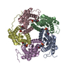
| ||||||||
|---|---|---|---|---|---|---|---|---|---|
| 1 |
| ||||||||
| Unit cell |
|
-Components
| #1: Protein | Mass: 18448.316 Da / Num. of mol.: 5 Source method: isolated from a genetically manipulated source Source: (gene. exp.) Plasmid: PNCO113 / Production host:  References: UniProt: P50861, 6,7-dimethyl-8-ribityllumazine synthase #2: Chemical | ChemComp-INJ /   Mass: 411.345 Da / Num. of mol.: 5 / Source method: obtained synthetically / Formula: C14H26N3O9P Mass: 411.345 Da / Num. of mol.: 5 / Source method: obtained synthetically / Formula: C14H26N3O9P#3: Water | ChemComp-HOH / |  Mass: 18.015 Da / Num. of mol.: 550 / Source method: isolated from a natural source / Formula: H2O Mass: 18.015 Da / Num. of mol.: 550 / Source method: isolated from a natural source / Formula: H2O |
|---|
-Experimental details
-Experiment
| Experiment | Method: X-RAY DIFFRACTION / Number of used crystals: 1 |
|---|
- Sample preparation
| Crystal | Density Matthews: 2.78 Å3/Da / Density % sol: 55.78 % | ||||||||||||||||||||
|---|---|---|---|---|---|---|---|---|---|---|---|---|---|---|---|---|---|---|---|---|---|
| Crystal grow | Temperature: 295 K / Method: vapor diffusion, sitting drop / pH: 7 Details: sodium phosphate, potassium phosphate, pH 7, VAPOR DIFFUSION, SITTING DROP, temperature 295K | ||||||||||||||||||||
| Crystal grow | *PLUS Temperature: 22 ℃ | ||||||||||||||||||||
| Components of the solutions | *PLUS
|
-Data collection
| Diffraction | Mean temperature: 105 K |
|---|---|
| Diffraction source | Source: SYNCHROTRON / Site: EMBL/DESY, HAMBURG  / Beamline: X11 / Wavelength: 0.908 / Beamline: X11 / Wavelength: 0.908 |
| Detector | Type: MARRESEARCH / Detector: IMAGE PLATE / Date: Aug 25, 1998 |
| Radiation | Protocol: SINGLE WAVELENGTH / Monochromatic (M) / Laue (L): M / Scattering type: x-ray |
| Radiation wavelength | Wavelength: 0.908 Å / Relative weight: 1 |
| Reflection | Resolution: 1.85→20 Å / Num. all: 90091 / Num. obs: 89967 / % possible obs: 99.7 % / Observed criterion σ(I): -3 / Redundancy: 15.71 % / Biso Wilson estimate: 19.19 Å2 / Rmerge(I) obs: 0.044 / Net I/σ(I): 32 |
| Reflection shell | Resolution: 1.85→1.87 Å / Rmerge(I) obs: 0.26 / % possible all: 99.6 |
| Reflection | *PLUS Lowest resolution: 9999 Å / Observed criterion σ(I): 1 / Num. measured all: 510352 |
| Reflection shell | *PLUS % possible obs: 99.6 % |
- Processing
| Software |
| |||||||||||||||||||||||||
|---|---|---|---|---|---|---|---|---|---|---|---|---|---|---|---|---|---|---|---|---|---|---|---|---|---|---|
| Refinement | Resolution: 1.85→20 Å / Stereochemistry target values: Engh & Huber
| |||||||||||||||||||||||||
| Displacement parameters | Biso mean: 22.094 Å2 | |||||||||||||||||||||||||
| Refinement step | Cycle: LAST / Resolution: 1.85→20 Å
| |||||||||||||||||||||||||
| Refine LS restraints |
| |||||||||||||||||||||||||
| Software | *PLUS Name: REFMAC / Classification: refinement | |||||||||||||||||||||||||
| Refinement | *PLUS % reflection Rfree: 5 % | |||||||||||||||||||||||||
| Solvent computation | *PLUS | |||||||||||||||||||||||||
| Displacement parameters | *PLUS | |||||||||||||||||||||||||
| Refine LS restraints | *PLUS
|
