 Movie
Movie Controller

[English] 日本語
 Yorodumi
- PDB-1c41: CRYSTAL STRUCTURES OF A PENTAMERIC FUNGAL AND AN ICOSAHEDRAL PLAN... -
+ Open data

- Basic information
| Entry | Database: PDB / ID: 1c41 | ||||||
|---|---|---|---|---|---|---|---|
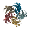
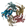
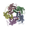
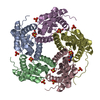
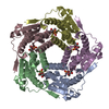
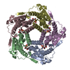
| Title | CRYSTAL STRUCTURES OF A PENTAMERIC FUNGAL AND AN ICOSAHEDRAL PLANT LUMAZINE SYNTHASE REVEALS THE STRUCTURAL BASIS FOR DIFFERENCES IN ASSEMBLY | ||||||
 Components Components | LUMAZINE SYNTHASE | ||||||
 Keywords Keywords | TRANSFERASE / RIBOFLAVIN BIOSYNTHESIS | ||||||
| Function / homology |  Function and homology information Function and homology information6,7-dimethyl-8-ribityllumazine synthase / 6,7-dimethyl-8-ribityllumazine synthase activity / riboflavin synthase complex / riboflavin biosynthetic process / mitochondrial intermembrane space Similarity search - Function | ||||||
| Biological species |  Magnaporthe grisea (fungus) Magnaporthe grisea (fungus) | ||||||
| Method |  X-RAY DIFFRACTION / SYNCHROTRON / MOLECULAR REPLACEMENT / Resolution: 3.1 Å X-RAY DIFFRACTION / SYNCHROTRON / MOLECULAR REPLACEMENT / Resolution: 3.1 Å | ||||||
 Authors Authors | Persson, K. / Schneider, G. / Jordan, D.B. / Viitanen, P.V. / Sandalova, T. | ||||||
 Citation Citation | Journal: Protein Sci. / Year: 1999 Title: Crystal structure analysis of a pentameric fungal and an icosahedral plant lumazine synthase reveals the structural basis for differences in assembly Authors: Persson, K. / Schneider, G. / Jordan, D.B. / Viitanen, P.V. / Sandalova, T. | ||||||
| History |
|
- Structure visualization
| Structure viewer | Molecule: MolmilJmol/JSmol |
|---|
- Downloads & links
-Download
| PDBx/mmCIF format | 1c41.cif.gz | 281.4 KB | Display | PDBx/mmCIF format |
|---|---|---|---|---|
| PDB format | pdb1c41.ent.gz | 223.8 KB | Display | PDB format |
| PDBx/mmJSON format | 1c41.json.gz | Tree view | PDBx/mmJSON format | |
| Others |  Other downloads Other downloads |
-Validation report
| Arichive directory | https://data.pdbj.org/pub/pdb/validation_reports/c4/1c41ftp://data.pdbj.org/pub/pdb/validation_reports/c4/1c41 | HTTPS FTP |
|---|
-Related structure data
| Related structure data | 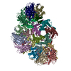 1c2yC 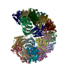 1rvvS S: Starting model for refinement C: citing same article ( |
|---|---|
| Similar structure data |


-Links
 PDBj
PDBj- Assembly
| Deposited unit | 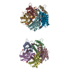
| ||||||||||||||||||||||||||||||||||||||||
|---|---|---|---|---|---|---|---|---|---|---|---|---|---|---|---|---|---|---|---|---|---|---|---|---|---|---|---|---|---|---|---|---|---|---|---|---|---|---|---|---|---|
| 1 | 
| ||||||||||||||||||||||||||||||||||||||||
| 2 | 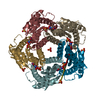
| ||||||||||||||||||||||||||||||||||||||||
| 3 | 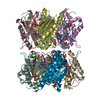
| ||||||||||||||||||||||||||||||||||||||||
| Unit cell |
| ||||||||||||||||||||||||||||||||||||||||
| Noncrystallographic symmetry (NCS) | NCS oper:
|
-Components
| #1: Protein | Mass: 21093.871 Da / Num. of mol.: 10 Source method: isolated from a genetically manipulated source Source: (gene. exp.) Magnaporthe grisea (fungus) / Production host:  References: UniProt: Q9UVT8, 6,7-dimethyl-8-ribityllumazine synthase #2: Chemical | ChemComp-SO4 /   Mass: 96.063 Da / Num. of mol.: 20 / Source method: obtained synthetically / Formula: SO4 Mass: 96.063 Da / Num. of mol.: 20 / Source method: obtained synthetically / Formula: SO4#3: Chemical | ChemComp-LMZ /   Mass: 290.230 Da / Num. of mol.: 10 / Source method: obtained synthetically / Formula: C9H14N4O7 Mass: 290.230 Da / Num. of mol.: 10 / Source method: obtained synthetically / Formula: C9H14N4O7#4: Water | ChemComp-HOH / |  Mass: 18.015 Da / Num. of mol.: 10 / Source method: isolated from a natural source / Formula: H2O Mass: 18.015 Da / Num. of mol.: 10 / Source method: isolated from a natural source / Formula: H2O |
|---|
-Experimental details
-Experiment
| Experiment | Method: X-RAY DIFFRACTION / Number of used crystals: 1 |
|---|
- Sample preparation
| Crystal | Density Matthews: 2.44 Å3/Da / Density % sol: 49.2 % | |||||||||||||||||||||||||
|---|---|---|---|---|---|---|---|---|---|---|---|---|---|---|---|---|---|---|---|---|---|---|---|---|---|---|
| Crystal grow | pH: 6.5 / Details: pH 6.5 | |||||||||||||||||||||||||
| Crystal grow | *PLUS Method: vapor diffusion, hanging drop | |||||||||||||||||||||||||
| Components of the solutions | *PLUS
|
-Data collection
| Diffraction | Mean temperature: 110 K |
|---|---|
| Diffraction source | Source: SYNCHROTRON / Site: MAX II  / Beamline: I711 / Wavelength: 0.958 / Beamline: I711 / Wavelength: 0.958 |
| Detector | Type: MARRESEARCH / Detector: IMAGE PLATE / Date: Nov 15, 1998 |
| Radiation | Protocol: SINGLE WAVELENGTH / Monochromatic (M) / Laue (L): M / Scattering type: x-ray |
| Radiation wavelength | Wavelength: 0.958 Å / Relative weight: 1 |
| Reflection | Resolution: 3.1→20 Å / Num. obs: 31842 / % possible obs: 99 % / Redundancy: 16.9 % / Rsym value: 0.136 / Net I/σ(I): 12 |
| Reflection shell | Resolution: 3.1→3.17 Å / Mean I/σ(I) obs: 3.3 / Rsym value: 0.396 / % possible all: 91.5 |
| Reflection | *PLUS Num. measured all: 539917 / Rmerge(I) obs: 0.136 |
| Reflection shell | *PLUS % possible obs: 91.5 % / Rmerge(I) obs: 0.396 |
- Processing
| Software |
| ||||||||||||||||||||||||||||||||||||||||||||||||||||||||||||
|---|---|---|---|---|---|---|---|---|---|---|---|---|---|---|---|---|---|---|---|---|---|---|---|---|---|---|---|---|---|---|---|---|---|---|---|---|---|---|---|---|---|---|---|---|---|---|---|---|---|---|---|---|---|---|---|---|---|---|---|---|---|
| Refinement | Method to determine structure: MOLECULAR REPLACEMENT Starting model: PDB ENTRY 1RVV Resolution: 3.1→20 Å / Cross valid method: THROUGHOUT / σ(F): 0
| ||||||||||||||||||||||||||||||||||||||||||||||||||||||||||||
| Displacement parameters | Biso mean: 23.3 Å2
| ||||||||||||||||||||||||||||||||||||||||||||||||||||||||||||
| Refinement step | Cycle: LAST / Resolution: 3.1→20 Å
| ||||||||||||||||||||||||||||||||||||||||||||||||||||||||||||
| Refine LS restraints |
| ||||||||||||||||||||||||||||||||||||||||||||||||||||||||||||
| Refine LS restraints NCS | NCS model details: STRICT | ||||||||||||||||||||||||||||||||||||||||||||||||||||||||||||
| LS refinement shell | Resolution: 3.1→3.13 Å / Total num. of bins used: 32
| ||||||||||||||||||||||||||||||||||||||||||||||||||||||||||||
| Xplor file |
|
