 Movie
Movie Controller


+ Open data

- Basic information
| Entry | Database: PDB / ID: 1zis | ||||||
|---|---|---|---|---|---|---|---|
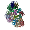
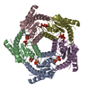
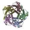
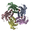
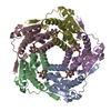
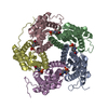
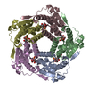
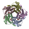
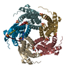
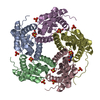
| Title | Recombinant Lumazine synthase (hexagonal form) | ||||||
 Components Components | 6,7-dimethyl-8-ribityllumazine synthase | ||||||
 Keywords Keywords | TRANSFERASE | ||||||
| Function / homology |  Function and homology information Function and homology information6,7-dimethyl-8-ribityllumazine synthase / 6,7-dimethyl-8-ribityllumazine synthase activity / riboflavin synthase complex / riboflavin biosynthetic process / cytoplasm / cytosol Similarity search - Function | ||||||
| Biological species |  | ||||||
| Method |  X-RAY DIFFRACTION / SYNCHROTRON / MOLECULAR REPLACEMENT / Resolution: 2.9 Å X-RAY DIFFRACTION / SYNCHROTRON / MOLECULAR REPLACEMENT / Resolution: 2.9 Å | ||||||
 Authors Authors | Lopez-Jaramillo, F.J. | ||||||
 Citation Citation | Journal: To be Published Title: Crystal structure of recombinant Lumazine synthase (hexagonal form) Authors: Lopez-Jaramillo, F.J. #1: Journal: J.Mol.Biol. / Year: 1995Title: Studies on the Lumazine Synthase/Riboflavin Synthase Complex of Bacillus subtilis: Crystal Structure Analysis of Reconstituted, Icosahedral b-subunit Capsids with Bound Substrate Analogue ...Title: Studies on the Lumazine Synthase/Riboflavin Synthase Complex of Bacillus subtilis: Crystal Structure Analysis of Reconstituted, Icosahedral b-subunit Capsids with Bound Substrate Analogue Inhibitor at 2.4 A Resolution Authors: Ritsert, K. / Huber, R. / Turk, D. / Ladenstein, R. / Scmidt-Base, K. / Bacher, A. #2: Journal: J.Mol.Biol. / Year: 1988Title: Heavy Riboflavin Synthase from Bacillus subtilis: Crystal Structure Analysis of the Icosahedral b60 Capsid at 3.3 A Resolution Authors: Ladenstein, R. / Schneider, M. / Huber, R. / Bartunik, H.-D. / Wilson, K. / Schott, K. / Bacher, A. #3: Journal: J.Biol.Chem. / Year: 1990Title: The Lumazine Synthase-Riboflavin Synthase Complex of Bacillus subtilis. Crystallization of Reconstituted Icosahedral b-subunit Capsids Authors: Schott, K. / Ladenstein, R. / Konig, A. / Bacher, A. | ||||||
| History |
|
- Structure visualization
| Structure viewer | Molecule: MolmilJmol/JSmol |
|---|
- Downloads & links
-Download
| PDBx/mmCIF format | 1zis.cif.gz | 267.6 KB | Display | PDBx/mmCIF format |
|---|---|---|---|---|
| PDB format | pdb1zis.ent.gz | 224.5 KB | Display | PDB format |
| PDBx/mmJSON format | 1zis.json.gz | Tree view | PDBx/mmJSON format | |
| Others |  Other downloads Other downloads |
-Validation report
| Arichive directory | https://data.pdbj.org/pub/pdb/validation_reports/zi/1zisftp://data.pdbj.org/pub/pdb/validation_reports/zi/1zis | HTTPS FTP |
|---|
-Related structure data
| Related structure data | 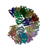 1rvvS S: Starting model for refinement |
|---|---|
| Similar structure data |


-Links
 PDBj
PDBj- Assembly
| Deposited unit | 
| ||||||||
|---|---|---|---|---|---|---|---|---|---|
| 1 | 
| ||||||||
| 2 | 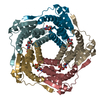
| ||||||||
| 3 | x 6 x 6 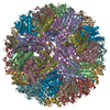
| ||||||||
| 4 | x 6 x 6 
| ||||||||
| Unit cell |
|
-Components
| #1: Protein | Mass: 16302.567 Da / Num. of mol.: 10 Source method: isolated from a genetically manipulated source Source: (gene. exp.) References: UniProt: P11998, 6,7-dimethyl-8-ribityllumazine synthase #2: Chemical | ChemComp-PO4 /   Mass: 94.971 Da / Num. of mol.: 10 / Source method: obtained synthetically / Formula: PO4 Mass: 94.971 Da / Num. of mol.: 10 / Source method: obtained synthetically / Formula: PO4#3: Chemical | ChemComp-INI /   Mass: 306.229 Da / Num. of mol.: 10 / Source method: obtained synthetically / Formula: C9H14N4O8 Mass: 306.229 Da / Num. of mol.: 10 / Source method: obtained synthetically / Formula: C9H14N4O8 |
|---|
-Experimental details
-Experiment
| Experiment | Method: X-RAY DIFFRACTION / Number of used crystals: 2 |
|---|
- Sample preparation
| Crystal | Density Matthews: 3 Å3/Da / Density % sol: 59.2 % |
|---|---|
| Crystal grow | Temperature: 293 K / Method: vapor diffusion, sitting drop / pH: 8.7 Details: Phosphate buffer, pH 8.7, VAPOR DIFFUSION, SITTING DROP, temperature 293K |
-Data collection
| Diffraction | Mean temperature: 100 K |
|---|---|
| Diffraction source | Source: SYNCHROTRON / Site: ESRF  / Beamline: BM16 / Wavelength: 0.98 Å / Beamline: BM16 / Wavelength: 0.98 Å |
| Detector | Type: MARRESEARCH / Detector: AREA DETECTOR / Date: Jun 22, 2004 / Details: mirrors |
| Radiation | Monochromator: Si 111 / Protocol: SINGLE WAVELENGTH / Monochromatic (M) / Laue (L): M / Scattering type: x-ray |
| Radiation wavelength | Wavelength: 0.98 Å / Relative weight: 1 |
| Reflection | Resolution: 2.9→19.6 Å / Num. obs: 40931 / % possible obs: 87.5 % / Observed criterion σ(F): 0 / Observed criterion σ(I): 0 / Redundancy: 7.3 % / Biso Wilson estimate: 44.616 Å2 / Limit h max: 45 / Limit h min: 0 / Limit k max: 26 / Limit k min: 0 / Limit l max: 102 / Limit l min: 0 / Observed criterion F max: 7253072.27 / Observed criterion F min: 83.39 / Rmerge(I) obs: 0.085 / Rsym value: 0.073 / Net I/σ(I): 17.59 |
| Reflection shell | Resolution: 2.9→3 Å / Redundancy: 4.3 % / Rmerge(I) obs: 0.19 / Mean I/σ(I) obs: 8.38 / Num. unique all: 3377 / Rsym value: 0.15 / % possible all: 77.1 |
- Processing
| Software |
| |||||||||||||||||||||||||
|---|---|---|---|---|---|---|---|---|---|---|---|---|---|---|---|---|---|---|---|---|---|---|---|---|---|---|
| Refinement | Method to determine structure: MOLECULAR REPLACEMENT Starting model: pentamer from 1RVV Resolution: 2.9→10 Å / Rfactor Rfree error: 0.004 / Occupancy max: 1 / Occupancy min: 1 / Cross valid method: THROUGHOUT / σ(F): 0 / σ(I): 0 / Stereochemistry target values: Engh & Huber
| |||||||||||||||||||||||||
| Solvent computation | Solvent model: CNS bulk solvent model used / Bsol: 24.9564 Å2 / ksol: 0.327727 e/Å3 | |||||||||||||||||||||||||
| Displacement parameters | Biso max: 194.39 Å2 / Biso mean: 44.29 Å2 / Biso min: 5.71 Å2
| |||||||||||||||||||||||||
| Refine analyze |
| |||||||||||||||||||||||||
| Refinement step | Cycle: LAST / Resolution: 2.9→10 Å
| |||||||||||||||||||||||||
| Refine LS restraints |
| |||||||||||||||||||||||||
| Xplor file |
|
