 Movie
Movie Controller


+ Open data

- Basic information
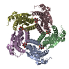
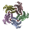
| Entry | Database: PDB / ID: 1kyv | ||||||
|---|---|---|---|---|---|---|---|
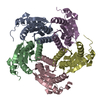
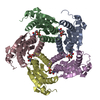
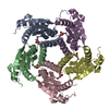
| Title | Lumazine Synthase from S.pombe bound to riboflavin | ||||||
 Components Components | 6,7-Dimethyl-8-ribityllumazine Synthase | ||||||
 Keywords Keywords | TRANSFERASE / riboflavin biosynthesis / lumazine synthase / Schizosaccharomyces pombe / ligand binding | ||||||
| Function / homology |  Function and homology information Function and homology informationriboflavin binding / 6,7-dimethyl-8-ribityllumazine synthase / 6,7-dimethyl-8-ribityllumazine synthase activity / riboflavin synthase complex / riboflavin biosynthetic process / mitochondrial intermembrane space / nucleus / cytoplasm Similarity search - Function | ||||||
| Biological species |  | ||||||
| Method |  X-RAY DIFFRACTION / MOLECULAR REPLACEMENT / Resolution: 2.4 Å X-RAY DIFFRACTION / MOLECULAR REPLACEMENT / Resolution: 2.4 Å | ||||||
 Authors Authors | Gerhardt, S. / Haase, I. / Steinbacher, S. / Kaiser, J.T. / Cushman, M. / Bacher, A. / Huber, R. / Fischer, M. | ||||||
 Citation Citation | Journal: J.Mol.Biol. / Year: 2002 Title: The structural basis of riboflavin binding to Schizosaccharomyces pombe 6,7-dimethyl-8-ribityllumazine synthase. Authors: Gerhardt, S. / Haase, I. / Steinbacher, S. / Kaiser, J.T. / Cushman, M. / Bacher, A. / Huber, R. / Fischer, M. | ||||||
| History |
|
- Structure visualization
| Structure viewer | Molecule: MolmilJmol/JSmol |
|---|
- Downloads & links
-Download
| PDBx/mmCIF format | 1kyv.cif.gz | 155.2 KB | Display | PDBx/mmCIF format |
|---|---|---|---|---|
| PDB format | pdb1kyv.ent.gz | 124.6 KB | Display | PDB format |
| PDBx/mmJSON format | 1kyv.json.gz | Tree view | PDBx/mmJSON format | |
| Others |  Other downloads Other downloads |
-Validation report
| Arichive directory | https://data.pdbj.org/pub/pdb/validation_reports/ky/1kyvftp://data.pdbj.org/pub/pdb/validation_reports/ky/1kyv | HTTPS FTP |
|---|
-Related structure data
| Related structure data |  1kyxC  1kyyC  1kz1C 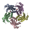 1kz4C 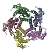 1kz6C  1kz9C C: citing same article ( |
|---|---|
| Similar structure data |


-Links
 PDBj
PDBj- Assembly
| Deposited unit | 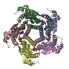
| ||||||||
|---|---|---|---|---|---|---|---|---|---|
| 1 |
| ||||||||
| 2 | 
| ||||||||
| Unit cell |
|
-Components
| #1: Protein | Mass: 17206.791 Da / Num. of mol.: 5 Source method: isolated from a genetically manipulated source Source: (gene. exp.) Plasmid: pNCO113 / Production host:  References: UniProt: Q9UUB1, 6,7-dimethyl-8-ribityllumazine synthase #2: Chemical | ChemComp-PO4 /   Mass: 94.971 Da / Num. of mol.: 5 / Source method: obtained synthetically / Formula: PO4 Mass: 94.971 Da / Num. of mol.: 5 / Source method: obtained synthetically / Formula: PO4#3: Chemical | ChemComp-RBF / 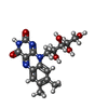  Mass: 376.364 Da / Num. of mol.: 5 / Source method: obtained synthetically / Formula: C17H20N4O6 Mass: 376.364 Da / Num. of mol.: 5 / Source method: obtained synthetically / Formula: C17H20N4O6#4: Water | ChemComp-HOH / |  Mass: 18.015 Da / Num. of mol.: 94 / Source method: isolated from a natural source / Formula: H2O Mass: 18.015 Da / Num. of mol.: 94 / Source method: isolated from a natural source / Formula: H2O |
|---|
-Experimental details
-Experiment
| Experiment | Method: X-RAY DIFFRACTION / Number of used crystals: 1 |
|---|
- Sample preparation
| Crystal | Density Matthews: 3.03 Å3/Da / Density % sol: 59.44 % | ||||||||||||||||||||||||||||||||||||||||||
|---|---|---|---|---|---|---|---|---|---|---|---|---|---|---|---|---|---|---|---|---|---|---|---|---|---|---|---|---|---|---|---|---|---|---|---|---|---|---|---|---|---|---|---|
| Crystal grow | Temperature: 295 K / Method: vapor diffusion, sitting drop / pH: 5.2 Details: citrate, sodium formate, pH 5.2, VAPOR DIFFUSION, SITTING DROP, temperature 295K | ||||||||||||||||||||||||||||||||||||||||||
| Crystal grow | *PLUS pH: 7 | ||||||||||||||||||||||||||||||||||||||||||
| Components of the solutions | *PLUS
|
-Data collection
| Diffraction | Mean temperature: 295 K |
|---|---|
| Diffraction source | Source: ROTATING ANODE / Type: RIGAKU RU200 / Wavelength: 1.5418 Å |
| Detector | Type: MARRESEARCH / Detector: IMAGE PLATE |
| Radiation | Monochromator: GRAPHITE / Protocol: SINGLE WAVELENGTH / Monochromatic (M) / Laue (L): M / Scattering type: x-ray |
| Radiation wavelength | Wavelength: 1.5418 Å / Relative weight: 1 |
| Reflection | Resolution: 2.4→16.03 Å / Num. all: 41051 / Num. obs: 40966 / % possible obs: 99.8 % / Observed criterion σ(F): 0 / Observed criterion σ(I): 0 |
| Reflection shell | Resolution: 2.4→2.53 Å / % possible all: 99.8 |
| Reflection | *PLUS % possible obs: 99.5 % / Redundancy: 3.5 % / Rmerge(I) obs: 0.078 |
| Reflection shell | *PLUS % possible obs: 99.5 % / Rmerge(I) obs: 0.45 |
- Processing
| Software |
| ||||||||||||||||||||
|---|---|---|---|---|---|---|---|---|---|---|---|---|---|---|---|---|---|---|---|---|---|
| Refinement | Method to determine structure: MOLECULAR REPLACEMENT / Resolution: 2.4→16.03 Å / σ(F): 0 / Stereochemistry target values: Engh & Huber
| ||||||||||||||||||||
| Refinement step | Cycle: LAST / Resolution: 2.4→16.03 Å
| ||||||||||||||||||||
| Refine LS restraints |
| ||||||||||||||||||||
| Refinement | *PLUS % reflection Rfree: 10 % / Rfactor all: 0.189 / Rfactor Rfree: 0.213 / Rfactor Rwork: 0.18 | ||||||||||||||||||||
| Solvent computation | *PLUS | ||||||||||||||||||||
| Displacement parameters | *PLUS | ||||||||||||||||||||
| Refine LS restraints | *PLUS
|
