 Movie
Movie Controller

[English] 日本語
 Yorodumi
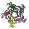
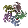
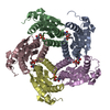
- PDB-2a57: Structure of 6,7-Dimthyl-8-ribityllumazine synthase from Schizosa... -
+ Open data

- Basic information
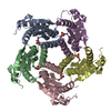
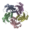
| Entry | Database: PDB / ID: 2a57 | ||||||
|---|---|---|---|---|---|---|---|
| Title | Structure of 6,7-Dimthyl-8-ribityllumazine synthase from Schizosaccharomyces pombe mutant W27Y with bound ligand 6-carboxyethyl-7-oxo-8-ribityllumazine | ||||||
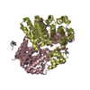
 Components Components | 6,7-dimethyl-8-ribityllumazine synthase | ||||||
 Keywords Keywords | TRANSFERASE / 6 / 7-dimethyl-8-ribityllumazine synthase / riboflavin / riboflavin biosynthesis / LUSY | ||||||
| Function / homology |  Function and homology information Function and homology informationriboflavin binding / 6,7-dimethyl-8-ribityllumazine synthase / 6,7-dimethyl-8-ribityllumazine synthase activity / riboflavin synthase complex / riboflavin biosynthetic process / mitochondrial intermembrane space / nucleus / cytoplasm Similarity search - Function | ||||||
| Biological species |  | ||||||
| Method |  X-RAY DIFFRACTION / FOURIER SYNTHESIS / Resolution: 2.75 Å X-RAY DIFFRACTION / FOURIER SYNTHESIS / Resolution: 2.75 Å | ||||||
 Authors Authors | Koch, M. / Breithaupt, C. / Gerhardt, S. / Haase, I. / Weber, S. / Cushman, M. / Huber, R. / Bacher, A. / Fischer, M. | ||||||
 Citation Citation | Journal: Eur.J.Biochem. / Year: 2004 Title: Structural basis of charge transfer complex formation by riboflavin bound to 6,7-dimethyl-8-ribityllumazine synthase Authors: Koch, M. / Breithaupt, C. / Gerhardt, S. / Haase, I. / Weber, S. / Cushman, M. / Huber, R. / Bacher, A. / Fischer, M. | ||||||
| History |
|
- Structure visualization
| Structure viewer | Molecule: MolmilJmol/JSmol |
|---|
- Downloads & links
-Download
| PDBx/mmCIF format | 2a57.cif.gz | 149.6 KB | Display | PDBx/mmCIF format |
|---|---|---|---|---|
| PDB format | pdb2a57.ent.gz | 119.1 KB | Display | PDB format |
| PDBx/mmJSON format | 2a57.json.gz | Tree view | PDBx/mmJSON format | |
| Others |  Other downloads Other downloads |
-Validation report
| Arichive directory | https://data.pdbj.org/pub/pdb/validation_reports/a5/2a57ftp://data.pdbj.org/pub/pdb/validation_reports/a5/2a57 | HTTPS FTP |
|---|
-Related structure data
| Related structure data |  2a58C 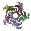 2a59C  1kyvS S: Starting model for refinement C: citing same article ( |
|---|---|
| Similar structure data |


-Links
 PDBj
PDBj- Assembly
| Deposited unit | 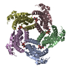
| ||||||||
|---|---|---|---|---|---|---|---|---|---|
| 1 |
| ||||||||
| 2 | 
| ||||||||
| Unit cell |
| ||||||||
| Details | the biological assembly is the pentamer found in the asymmetric unit |
-Components
| #1: Protein | Mass: 17183.756 Da / Num. of mol.: 5 / Mutation: W27Y Source method: isolated from a genetically manipulated source Source: (gene. exp.) Gene: rib4 / Production host:  References: UniProt: Q9UUB1, 6,7-dimethyl-8-ribityllumazine synthase #2: Chemical | ChemComp-CRM /   Mass: 386.314 Da / Num. of mol.: 5 / Source method: obtained synthetically / Formula: C14H18N4O9 Mass: 386.314 Da / Num. of mol.: 5 / Source method: obtained synthetically / Formula: C14H18N4O9#3: Chemical | ChemComp-PO4 /   Mass: 94.971 Da / Num. of mol.: 5 / Source method: obtained synthetically / Formula: PO4 Mass: 94.971 Da / Num. of mol.: 5 / Source method: obtained synthetically / Formula: PO4 |
|---|
-Experimental details
-Experiment
| Experiment | Method: X-RAY DIFFRACTION / Number of used crystals: 1 |
|---|
- Sample preparation
| Crystal | Density Matthews: 2.87 Å3/Da / Density % sol: 57.1 % |
|---|---|
| Crystal grow | Temperature: 291 K / Method: vapor diffusion, sitting drop / pH: 5 Details: sodium citrate, ammonium dihydrogen phosphate, pH 5.0, VAPOR DIFFUSION, SITTING DROP, temperature 291K |
-Data collection
| Diffraction | Mean temperature: 298 K |
|---|---|
| Diffraction source | Source: ROTATING ANODE / Type: RIGAKU RU200 / Wavelength: 1.5418 Å |
| Detector | Type: MAR scanner 345 mm plate / Detector: IMAGE PLATE / Date: Jul 17, 2002 |
| Radiation | Protocol: SINGLE WAVELENGTH / Monochromatic (M) / Laue (L): M / Scattering type: x-ray |
| Radiation wavelength | Wavelength: 1.5418 Å / Relative weight: 1 |
| Reflection | Resolution: 2.75→20 Å / Num. all: 26937 / Num. obs: 26937 / % possible obs: 99.4 % / Observed criterion σ(F): 2 / Observed criterion σ(I): 2 / Redundancy: 3.9 % / Rmerge(I) obs: 0.08 / Rsym value: 0.08 / Net I/σ(I): 15.5 |
| Reflection shell | Resolution: 2.75→2.83 Å / Redundancy: 3.8 % / Rmerge(I) obs: 0.517 / Mean I/σ(I) obs: 2.6 / Rsym value: 0.517 / % possible all: 99.5 |
- Processing
| Software |
| ||||||||||||||||||||||||||||
|---|---|---|---|---|---|---|---|---|---|---|---|---|---|---|---|---|---|---|---|---|---|---|---|---|---|---|---|---|---|
| Refinement | Method to determine structure: FOURIER SYNTHESIS Starting model: PDB ENTRY 1KYV Resolution: 2.75→25 Å / Isotropic thermal model: Isotropic / σ(F): 0 / Stereochemistry target values: Engh & Huber
| ||||||||||||||||||||||||||||
| Solvent computation | Bsol: 58.754 Å2 | ||||||||||||||||||||||||||||
| Displacement parameters | Biso mean: 61.234 Å2
| ||||||||||||||||||||||||||||
| Refinement step | Cycle: LAST / Resolution: 2.75→25 Å
| ||||||||||||||||||||||||||||
| Refine LS restraints |
| ||||||||||||||||||||||||||||
| Xplor file |
|
