 Movie
Movie Controller


+ Open data

- Basic information
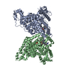
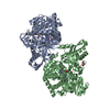
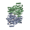
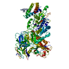
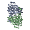
| Entry | Database: PDB / ID: 1e4e | ||||||
|---|---|---|---|---|---|---|---|
| Title | D-alanyl-D-lacate ligase | ||||||
 Components Components | (VANCOMYCIN/TEICOPLANIN A-TYPE RESISTANCE PROTEIN ...) x 2 | ||||||
 Keywords Keywords | LIGASE / CELL WALL / ANTIBIOTIC RESISTANCE / MEMBRANE / PEPTIDOGLYCAN SYNTHESIS | ||||||
| Function / homology |  Function and homology information Function and homology informationD-alanine-(R)-lactate ligase / D-alanine-(R)-lactate ligase activity / D-alanine-D-alanine ligase activity / peptidoglycan biosynthetic process / cell wall organization / regulation of cell shape / response to antibiotic / ATP binding / metal ion binding / plasma membrane / cytosol Similarity search - Function | ||||||
| Biological species |  ENTEROCOCCUS FAECIUM (bacteria) ENTEROCOCCUS FAECIUM (bacteria) | ||||||
| Method |  X-RAY DIFFRACTION / MOLECULAR REPLACEMENT / Resolution: 2.5 Å X-RAY DIFFRACTION / MOLECULAR REPLACEMENT / Resolution: 2.5 Å | ||||||
 Authors Authors | Roper, D.I. | ||||||
 Citation Citation | Journal: Proc. Natl. Acad. Sci. U.S.A. / Year: 2000 Title: The molecular basis of vancomycin resistance in clinically relevant Enterococci: crystal structure of D-alanyl-D-lactate ligase (VanA). Authors: Roper, D.I. / Huyton, T. / Vagin, A. / Dodson, G. | ||||||
| History |
|
- Structure visualization
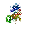
| Structure viewer | Molecule: MolmilJmol/JSmol |
|---|
- Downloads & links
-Download
| PDBx/mmCIF format | 1e4e.cif.gz | 159.6 KB | Display | PDBx/mmCIF format |
|---|---|---|---|---|
| PDB format | pdb1e4e.ent.gz | 123.9 KB | Display | PDB format |
| PDBx/mmJSON format | 1e4e.json.gz | Tree view | PDBx/mmJSON format | |
| Others |  Other downloads Other downloads |
-Validation report
| Arichive directory | https://data.pdbj.org/pub/pdb/validation_reports/e4/1e4eftp://data.pdbj.org/pub/pdb/validation_reports/e4/1e4e | HTTPS FTP |
|---|
-Related structure data
| Related structure data | 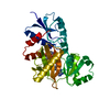 1iowS S: Starting model for refinement |
|---|---|
| Similar structure data |

-Links
 PDBj
PDBj


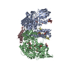
- Assembly
| Deposited unit | 
| ||||||||
|---|---|---|---|---|---|---|---|---|---|
| 1 | 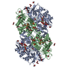
| ||||||||
| Unit cell |
| ||||||||
| Noncrystallographic symmetry (NCS) | NCS oper: (Code: given Matrix: (-0.57983, -0.49666, -0.64586), Vector: |
-Components
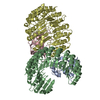
-VANCOMYCIN/TEICOPLANIN A-TYPE RESISTANCE PROTEIN ... , 2 types, 2 molecules AB
| #1: Protein | Mass: 37575.047 Da / Num. of mol.: 1 Source method: isolated from a genetically manipulated source Source: (gene. exp.) ENTEROCOCCUS FAECIUM (bacteria) / Strain: BM41417 / Cellular location: CYTOPLASM / Gene: VANA / Production host: |
|---|---|
| #2: Protein | Mass: 37618.141 Da / Num. of mol.: 1 Source method: isolated from a genetically manipulated source Source: (gene. exp.) ENTEROCOCCUS FAECIUM (bacteria) / Strain: BM41417 / Cellular location: CYTOPLASM / Gene: VANA / Production host: |
-Non-polymers , 6 types, 380 molecules 
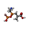


| #3: Chemical |  Mass: 427.201 Da / Num. of mol.: 2 / Source method: obtained synthetically / Formula: C10H15N5O10P2 / Comment: ADP, energy-carrying molecule*YM Mass: 427.201 Da / Num. of mol.: 2 / Source method: obtained synthetically / Formula: C10H15N5O10P2 / Comment: ADP, energy-carrying molecule*YM#4: Chemical |  Mass: 275.133 Da / Num. of mol.: 2 / Source method: obtained synthetically / Formula: C6H15NO7P2 Mass: 275.133 Da / Num. of mol.: 2 / Source method: obtained synthetically / Formula: C6H15NO7P2#5: Chemical | ChemComp-MG /  Mass: 24.305 Da / Num. of mol.: 4 / Source method: obtained synthetically / Formula: Mg Mass: 24.305 Da / Num. of mol.: 4 / Source method: obtained synthetically / Formula: Mg#6: Chemical |  Mass: 96.063 Da / Num. of mol.: 3 / Source method: obtained synthetically / Formula: SO4 Mass: 96.063 Da / Num. of mol.: 3 / Source method: obtained synthetically / Formula: SO4#7: Chemical | ChemComp-GOL /  Mass: 92.094 Da / Num. of mol.: 14 / Source method: obtained synthetically / Formula: C3H8O3 Mass: 92.094 Da / Num. of mol.: 14 / Source method: obtained synthetically / Formula: C3H8O3#8: Water | ChemComp-HOH / | Mass: 18.015 Da / Num. of mol.: 355 / Source method: isolated from a natural source / Formula: H2O |
|---|
-Details
| Has protein modification | Y |
|---|---|
| Sequence details | CHAIN A AND CHAIN B DIFFER AT RESIDUE 298 |
-Experimental details
-Experiment
| Experiment | Method: X-RAY DIFFRACTION |
|---|
- Sample preparation
| Crystal | Density Matthews: 3.36 Å3/Da / Density % sol: 63.13 % | ||||||||||||||||||
|---|---|---|---|---|---|---|---|---|---|---|---|---|---|---|---|---|---|---|---|
| Crystal grow | Method: vapor diffusion, hanging drop / pH: 6.5 / Details: pH 6.50 | ||||||||||||||||||
| Crystal grow | *PLUS pH: 6 / Method: vapor diffusion, hanging drop | ||||||||||||||||||
| Components of the solutions | *PLUS
|
-Data collection
| Diffraction | Mean temperature: 120 K |
|---|---|
| Diffraction source | Source: ROTATING ANODE / Wavelength: 1.5418 |
| Detector | Date: Jan 15, 1998 |
| Radiation | Protocol: SINGLE WAVELENGTH / Monochromatic (M) / Laue (L): M / Scattering type: x-ray |
| Radiation wavelength | Wavelength: 1.5418 Å / Relative weight: 1 |
| Reflection | Resolution: 2.5→15 Å / Num. obs: 32814 / % possible obs: 93.3 % / Observed criterion σ(I): 2 / Redundancy: 1 % / Biso Wilson estimate: 41.66 Å2 / Rmerge(I) obs: 0.058 / Net I/σ(I): 29.9 |
| Reflection shell | Resolution: 2.5→2.54 Å / Rmerge(I) obs: 0.217 / Mean I/σ(I) obs: 4.1 / % possible all: 96.6 |
| Reflection shell | *PLUS Highest resolution: 2.5 Å / % possible obs: 96.6 % / Rmerge(I) obs: 0.217 / Mean I/σ(I) obs: 4.1 |
- Processing
| Software |
| ||||||||||||||||||||||||||||||||||||||||||||||||||||||||||||||||||||||||||||||||||||
|---|---|---|---|---|---|---|---|---|---|---|---|---|---|---|---|---|---|---|---|---|---|---|---|---|---|---|---|---|---|---|---|---|---|---|---|---|---|---|---|---|---|---|---|---|---|---|---|---|---|---|---|---|---|---|---|---|---|---|---|---|---|---|---|---|---|---|---|---|---|---|---|---|---|---|---|---|---|---|---|---|---|---|---|---|---|
| Refinement | Method to determine structure: MOLECULAR REPLACEMENT Starting model: PDB ENTRY 1IOW Resolution: 2.5→15 Å / SU B: 0.191 / SU ML: 0.189 / Cross valid method: THROUGHOUT / σ(F): 1 / ESU R: 0.39 / ESU R Free: 0.29
| ||||||||||||||||||||||||||||||||||||||||||||||||||||||||||||||||||||||||||||||||||||
| Displacement parameters | Biso mean: 44.8 Å2
| ||||||||||||||||||||||||||||||||||||||||||||||||||||||||||||||||||||||||||||||||||||
| Refinement step | Cycle: LAST / Resolution: 2.5→15 Å
| ||||||||||||||||||||||||||||||||||||||||||||||||||||||||||||||||||||||||||||||||||||
| Refine LS restraints |
| ||||||||||||||||||||||||||||||||||||||||||||||||||||||||||||||||||||||||||||||||||||
| Software | *PLUS Name: REFMAC / Classification: refinement | ||||||||||||||||||||||||||||||||||||||||||||||||||||||||||||||||||||||||||||||||||||
| Refinement | *PLUS Lowest resolution: 15 Å / Rfactor obs: 0.183 / Rfactor Rfree: 0.284 / Rfactor Rwork: 0.205 | ||||||||||||||||||||||||||||||||||||||||||||||||||||||||||||||||||||||||||||||||||||
| Solvent computation | *PLUS | ||||||||||||||||||||||||||||||||||||||||||||||||||||||||||||||||||||||||||||||||||||
| Displacement parameters | *PLUS | ||||||||||||||||||||||||||||||||||||||||||||||||||||||||||||||||||||||||||||||||||||
| Refine LS restraints | *PLUS
|
