 Movie
Movie Controller

[English] 日本語
 Yorodumi
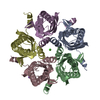
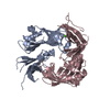
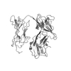
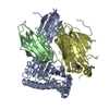
- PDB-6m8r: Crystal structure of the KCTD16 BTB domain in complex with GABAB2... -
+ Open data

- Basic information
| Entry | Database: PDB / ID: 6m8r | ||||||
|---|---|---|---|---|---|---|---|
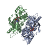
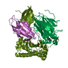
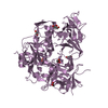
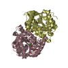
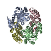
| Title | Crystal structure of the KCTD16 BTB domain in complex with GABAB2 peptide | ||||||
 Components Components |
| ||||||
 Keywords Keywords | PROTEIN BINDING / pentamer / BTB domain | ||||||
| Function / homology |  Function and homology information Function and homology informationGABA B receptor activation / G protein-coupled GABA receptor complex / : / neuron-glial cell signaling / G protein-coupled receptor heterodimeric complex / G protein-coupled GABA receptor activity / : / GABA receptor complex / negative regulation of adenylate cyclase activity / regulation of G protein-coupled receptor signaling pathway ...GABA B receptor activation / G protein-coupled GABA receptor complex / : / neuron-glial cell signaling / G protein-coupled receptor heterodimeric complex / G protein-coupled GABA receptor activity / : / GABA receptor complex / negative regulation of adenylate cyclase activity / regulation of G protein-coupled receptor signaling pathway / Class C/3 (Metabotropic glutamate/pheromone receptors) / gamma-aminobutyric acid signaling pathway / synaptic transmission, GABAergic / behavioral fear response / protein homooligomerization / memory / adenylate cyclase-inhibiting G protein-coupled receptor signaling pathway / Activation of G protein gated Potassium channels / Inhibition of voltage gated Ca2+ channels via Gbeta/gamma subunits / transmembrane signaling receptor activity / presynaptic membrane / chemical synaptic transmission / G alpha (i) signalling events / postsynaptic membrane / signaling receptor complex / neuron projection / G protein-coupled receptor signaling pathway / protein heterodimerization activity / identical protein binding / plasma membrane / cytoplasm Similarity search - Function | ||||||
| Biological species |  Homo sapiens (human) Homo sapiens (human) | ||||||
| Method |  X-RAY DIFFRACTION / SYNCHROTRON / MOLECULAR REPLACEMENT / Resolution: 3.2 Å X-RAY DIFFRACTION / SYNCHROTRON / MOLECULAR REPLACEMENT / Resolution: 3.2 Å | ||||||
 Authors Authors | Zheng, S. / Kruse, A.C. | ||||||
| Funding support |  United States, 1items United States, 1items
| ||||||
 Citation Citation | Journal: Nature / Year: 2019 Title: Structural basis for KCTD-mediated rapid desensitization of GABABsignalling. Authors: Zheng, S. / Abreu, N. / Levitz, J. / Kruse, A.C. | ||||||
| History |
|
- Structure visualization
| Structure viewer | Molecule: MolmilJmol/JSmol |
|---|
- Downloads & links
-Download
| PDBx/mmCIF format | 6m8r.cif.gz | 220.1 KB | Display | PDBx/mmCIF format |
|---|---|---|---|---|
| PDB format | pdb6m8r.ent.gz | 177.9 KB | Display | PDB format |
| PDBx/mmJSON format | 6m8r.json.gz | Tree view | PDBx/mmJSON format | |
| Others |  Other downloads Other downloads |
-Validation report
| Arichive directory | https://data.pdbj.org/pub/pdb/validation_reports/m8/6m8rftp://data.pdbj.org/pub/pdb/validation_reports/m8/6m8r | HTTPS FTP |
|---|
-Related structure data
| Related structure data |  6m8sC 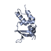 5a15S C: citing same article ( S: Starting model for refinement |
|---|---|
| Similar structure data |

-Links
 PDBj
PDBj


- Assembly
| Deposited unit | 
| ||||||||
|---|---|---|---|---|---|---|---|---|---|
| 1 | 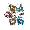
| ||||||||
| 2 | 
| ||||||||
| Unit cell |
|
-Components
| #1: Protein | Mass: 12220.125 Da / Num. of mol.: 10 / Fragment: UNP residues 23-124 Source method: isolated from a genetically manipulated source Source: (gene. exp.) Homo sapiens (human) / Gene: KCTD16, KIAA1317Production host:  References: UniProt: Q68DU8 #2: Protein/peptide | Mass: 4520.067 Da / Num. of mol.: 2 Source method: isolated from a genetically manipulated source Source: (gene. exp.) Homo sapiens (human) / Gene: GABBR2, GPR51, GPRC3BProduction host: References: UniProt: O75899 #3: Chemical |   Mass: 24.305 Da / Num. of mol.: 2 / Source method: obtained synthetically / Formula: Mg Mass: 24.305 Da / Num. of mol.: 2 / Source method: obtained synthetically / Formula: Mg |
|---|
-Experimental details
-Experiment
| Experiment | Method: X-RAY DIFFRACTION / Number of used crystals: 1 |
|---|
- Sample preparation
| Crystal | Density Matthews: 2.72 Å3/Da / Density % sol: 54.84 % |
|---|---|
| Crystal grow | Temperature: 293 K / Method: vapor diffusion, sitting drop Details: 100 mM magnesium chloride, 100 mM Tris-HCl, pH 7.5, 12% w/v PEG8000 |
-Data collection
| Diffraction | Mean temperature: 80 K |
|---|---|
| Diffraction source | Source: SYNCHROTRON / Site: APS / Beamline: 23-ID-B / Wavelength: 1.033 Å |
| Detector | Type: DECTRIS EIGER X 16M / Detector: PIXEL / Date: Jun 7, 2017 |
| Radiation | Monochromator: double crystal cryo-cooled Si(111) / Protocol: SINGLE WAVELENGTH / Monochromatic (M) / Laue (L): M / Scattering type: x-ray |
| Radiation wavelength | Wavelength: 1.033 Å / Relative weight: 1 |
| Reflection | Resolution: 3.2→50 Å / Num. obs: 22162 / % possible obs: 99.2 % / Redundancy: 3.3 % / CC1/2: 0.926 / Rmerge(I) obs: 0.332 / Rpim(I) all: 0.208 / Rrim(I) all: 0.394 / Net I/σ(I): 3.4 |
| Reflection shell | Resolution: 3.2→3.26 Å / Redundancy: 2.7 % / Rmerge(I) obs: 0.997 / Mean I/σ(I) obs: 0.6 / Num. unique obs: 1073 / CC1/2: 0.561 / Rpim(I) all: 0.703 / Rrim(I) all: 1.226 / % possible all: 96.7 |
- Processing
| Software |
| |||||||||||||||||||||||||||||||||||||||||||||||||||||||||||||||||||||||||||||||||||||||||||
|---|---|---|---|---|---|---|---|---|---|---|---|---|---|---|---|---|---|---|---|---|---|---|---|---|---|---|---|---|---|---|---|---|---|---|---|---|---|---|---|---|---|---|---|---|---|---|---|---|---|---|---|---|---|---|---|---|---|---|---|---|---|---|---|---|---|---|---|---|---|---|---|---|---|---|---|---|---|---|---|---|---|---|---|---|---|---|---|---|---|---|---|---|
| Refinement | Method to determine structure: MOLECULAR REPLACEMENT Starting model: PDB entry 5A15 Resolution: 3.2→38.639 Å / SU ML: 0.46 / Cross valid method: FREE R-VALUE / σ(F): 1.33 / Phase error: 27.73 / Stereochemistry target values: ML
| |||||||||||||||||||||||||||||||||||||||||||||||||||||||||||||||||||||||||||||||||||||||||||
| Solvent computation | Shrinkage radii: 0.9 Å / VDW probe radii: 1.11 Å / Solvent model: FLAT BULK SOLVENT MODEL | |||||||||||||||||||||||||||||||||||||||||||||||||||||||||||||||||||||||||||||||||||||||||||
| Refinement step | Cycle: LAST / Resolution: 3.2→38.639 Å
| |||||||||||||||||||||||||||||||||||||||||||||||||||||||||||||||||||||||||||||||||||||||||||
| Refine LS restraints |
| |||||||||||||||||||||||||||||||||||||||||||||||||||||||||||||||||||||||||||||||||||||||||||
| LS refinement shell |
|
