 Movie
Movie Controller

[English] 日本語
 Yorodumi
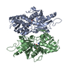
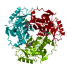
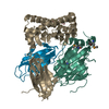
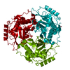
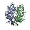
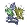
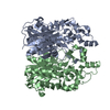
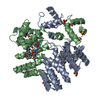
- PDB-5iwc: Mycobacterium tuberculosis CysM in complex with the Urea-scaffold... -
+ Open data

- Basic information
| Entry | Database: PDB / ID: 5iwc | ||||||
|---|---|---|---|---|---|---|---|
| Title | Mycobacterium tuberculosis CysM in complex with the Urea-scaffold inhibitor 3 [4-(3-([1,1'-Biphenyl]-3-yl)ureido)-2-hydroxybenzoic acid] | ||||||
 Components Components | O-phosphoserine sulfhydrylase | ||||||
 Keywords Keywords | LYASE / Mycobacterium tuberculosis / Cysteine biosynthesis / sulphur metabolism / inhibitor / transferase | ||||||
| Function / homology |  Function and homology information Function and homology information[CysO sulfur-carrier protein]-thiocarboxylate-dependent cysteine synthase / Cysteine synthesis from O-phosphoserine / O-phosphoserine sulfhydrylase activity / L-cysteine biosynthetic process / cysteine synthase activity / : / pyridoxal phosphate binding / protein-containing complex / cytoplasm / cytosol Similarity search - Function | ||||||
| Biological species |   Mycobacterium tuberculosis (bacteria) Mycobacterium tuberculosis (bacteria) | ||||||
| Method |  X-RAY DIFFRACTION / SYNCHROTRON / MOLECULAR REPLACEMENT / Resolution: 2.7 Å X-RAY DIFFRACTION / SYNCHROTRON / MOLECULAR REPLACEMENT / Resolution: 2.7 Å | ||||||
 Authors Authors | Schnell, R. / Maric, S. / Lindqvist, Y. / Schneider, G. | ||||||
 Citation Citation | Journal: J.Med.Chem. / Year: 2016 Title: Inhibitors of the Cysteine Synthase CysM with Antibacterial Potency against Dormant Mycobacterium tuberculosis. Authors: Brunner, K. / Maric, S. / Reshma, R.S. / Almqvist, H. / Seashore-Ludlow, B. / Gustavsson, A.L. / Poyraz, O. / Yogeeswari, P. / Lundback, T. / Vallin, M. / Sriram, D. / Schnell, R. / Schneider, G. | ||||||
| History |
|
- Structure visualization
| Structure viewer | Molecule: MolmilJmol/JSmol |
|---|
- Downloads & links
-Download
| PDBx/mmCIF format | 5iwc.cif.gz | 127.8 KB | Display | PDBx/mmCIF format |
|---|---|---|---|---|
| PDB format | pdb5iwc.ent.gz | 98 KB | Display | PDB format |
| PDBx/mmJSON format | 5iwc.json.gz | Tree view | PDBx/mmJSON format | |
| Others |  Other downloads Other downloads |
-Validation report
| Arichive directory | https://data.pdbj.org/pub/pdb/validation_reports/iw/5iwcftp://data.pdbj.org/pub/pdb/validation_reports/iw/5iwc | HTTPS FTP |
|---|
-Related structure data
| Related structure data | 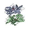 5i6dC 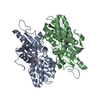 5i7aC 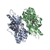 5i7hC 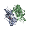 5i7oC 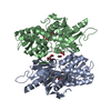 5i7rC 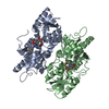 5iw8C 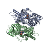 3dkiS S: Starting model for refinement C: citing same article ( |
|---|---|
| Similar structure data |


-Links
 PDBj
PDBj
- Assembly
| Deposited unit | 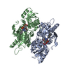
| ||||||||||||||||||
|---|---|---|---|---|---|---|---|---|---|---|---|---|---|---|---|---|---|---|---|
| 1 |
| ||||||||||||||||||
| Unit cell |
| ||||||||||||||||||
| Noncrystallographic symmetry (NCS) | NCS domain:
NCS domain segments: Component-ID: _ / Ens-ID: 1 / Beg auth comp-ID: ALA / Beg label comp-ID: ALA / End auth comp-ID: LEU / End label comp-ID: LEU / Refine code: _ / Auth seq-ID: 2 - 317 / Label seq-ID: 5 - 320
|
-Components
| #1: Protein | Mass: 34724.117 Da / Num. of mol.: 2 / Mutation: T2A Source method: isolated from a genetically manipulated source Details: Mutation: T2A, Lys51 covalent modification with PLP Source: (gene. exp.) Mycobacterium tuberculosis (strain ATCC 25618 / H37Rv) (bacteria)Gene: cysM, Rv1336, MTCY130.21 / Plasmid: pET28a Details (production host): N-terminal His6-tah, Thrombin site for tag-removal Production host: References: UniProt: P9WP53, [CysO sulfur-carrier protein]-thiocarboxylate-dependent cysteine synthase #2: Chemical |   Mass: 247.142 Da / Num. of mol.: 2 / Source method: obtained synthetically / Formula: C8H10NO6P Mass: 247.142 Da / Num. of mol.: 2 / Source method: obtained synthetically / Formula: C8H10NO6P#3: Chemical | 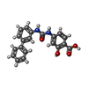  Mass: 348.352 Da / Num. of mol.: 2 / Source method: obtained synthetically / Formula: C20H16N2O4 Mass: 348.352 Da / Num. of mol.: 2 / Source method: obtained synthetically / Formula: C20H16N2O4#4: Water | ChemComp-HOH / |  Mass: 18.015 Da / Num. of mol.: 36 / Source method: isolated from a natural source / Formula: H2O Mass: 18.015 Da / Num. of mol.: 36 / Source method: isolated from a natural source / Formula: H2O |
|---|
-Experimental details
-Experiment
| Experiment | Method: X-RAY DIFFRACTION / Number of used crystals: 1 |
|---|
- Sample preparation
| Crystal | Density Matthews: 1.93 Å3/Da / Density % sol: 36.15 % / Description: rod / needle |
|---|---|
| Crystal grow | Temperature: 293 K / Method: vapor diffusion / pH: 5.5 Details: 0.1M Bis-Tris pH 5.5, 0.2M NH4-acetate, 25% PEG 3350 |
-Data collection
| Diffraction | Mean temperature: 100 K |
|---|---|
| Diffraction source | Source: SYNCHROTRON / Site: ESRF  / Beamline: ID23-1 / Wavelength: 0.97242 Å / Beamline: ID23-1 / Wavelength: 0.97242 Å |
| Detector | Type: DECTRIS PILATUS 6M / Detector: PIXEL / Date: Jun 18, 2014 / Details: mirrors |
| Radiation | Monochromator: Si 111 / Protocol: SINGLE WAVELENGTH / Monochromatic (M) / Laue (L): M / Scattering type: x-ray |
| Radiation wavelength | Wavelength: 0.97242 Å / Relative weight: 1 |
| Reflection | Resolution: 2.7→66.04 Å / Num. obs: 15620 / % possible obs: 96.9 % / Observed criterion σ(I): 2.1 / Redundancy: 5 % / Biso Wilson estimate: 39.1 Å2 / Rmerge(I) obs: 0.131 / Net I/σ(I): 9.1 |
| Reflection shell | Resolution: 2.7→2.85 Å / Redundancy: 4.9 % / Rmerge(I) obs: 0.806 / Mean I/σ(I) obs: 2.1 / % possible all: 95.2 |
- Processing
| Software |
| ||||||||||||||||||||||||||||||||||||||||||||||||||||||||||||||||||||||||||||||||||||||||||||||||||||||||||||||||||||||||||||||||||||||||||||||||||||||||||||||||||||||||||||||||||||||
|---|---|---|---|---|---|---|---|---|---|---|---|---|---|---|---|---|---|---|---|---|---|---|---|---|---|---|---|---|---|---|---|---|---|---|---|---|---|---|---|---|---|---|---|---|---|---|---|---|---|---|---|---|---|---|---|---|---|---|---|---|---|---|---|---|---|---|---|---|---|---|---|---|---|---|---|---|---|---|---|---|---|---|---|---|---|---|---|---|---|---|---|---|---|---|---|---|---|---|---|---|---|---|---|---|---|---|---|---|---|---|---|---|---|---|---|---|---|---|---|---|---|---|---|---|---|---|---|---|---|---|---|---|---|---|---|---|---|---|---|---|---|---|---|---|---|---|---|---|---|---|---|---|---|---|---|---|---|---|---|---|---|---|---|---|---|---|---|---|---|---|---|---|---|---|---|---|---|---|---|---|---|---|---|
| Refinement | Method to determine structure: MOLECULAR REPLACEMENT Starting model: 3dki Resolution: 2.7→65.43 Å / Cor.coef. Fo:Fc: 0.933 / Cor.coef. Fo:Fc free: 0.889 / SU B: 15.982 / SU ML: 0.327 / Cross valid method: THROUGHOUT / ESU R Free: 0.412 / Details: HYDROGENS HAVE BEEN ADDED IN THE RIDING POSITIONS
| ||||||||||||||||||||||||||||||||||||||||||||||||||||||||||||||||||||||||||||||||||||||||||||||||||||||||||||||||||||||||||||||||||||||||||||||||||||||||||||||||||||||||||||||||||||||
| Solvent computation | Ion probe radii: 0.8 Å / Shrinkage radii: 0.8 Å / VDW probe radii: 1.2 Å | ||||||||||||||||||||||||||||||||||||||||||||||||||||||||||||||||||||||||||||||||||||||||||||||||||||||||||||||||||||||||||||||||||||||||||||||||||||||||||||||||||||||||||||||||||||||
| Displacement parameters | Biso mean: 57.24 Å2
| ||||||||||||||||||||||||||||||||||||||||||||||||||||||||||||||||||||||||||||||||||||||||||||||||||||||||||||||||||||||||||||||||||||||||||||||||||||||||||||||||||||||||||||||||||||||
| Refinement step | Cycle: 1 / Resolution: 2.7→65.43 Å
| ||||||||||||||||||||||||||||||||||||||||||||||||||||||||||||||||||||||||||||||||||||||||||||||||||||||||||||||||||||||||||||||||||||||||||||||||||||||||||||||||||||||||||||||||||||||
| Refine LS restraints |
|