 Movie
Movie Controller

[English] 日本語
 Yorodumi
- PDB-6ns5: Crystal structure of fungal lipoxygenase from Fusarium graminearu... -
+ Open data

- Basic information
| Entry | Database: PDB / ID: 6ns5 | |||||||||
|---|---|---|---|---|---|---|---|---|---|---|
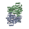
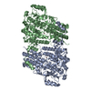
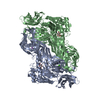
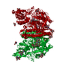
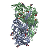
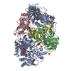
| Title | Crystal structure of fungal lipoxygenase from Fusarium graminearum. Second C2 crystal form. | |||||||||
 Components Components | lipoxygenase | |||||||||
 Keywords Keywords | OXIDOREDUCTASE / lipoxygenase / fungus / Fe coordination | |||||||||
| Function / homology |  Function and homology information Function and homology informationlinoleate 11-lipoxygenase / linoleate 11-lipoxygenase activity / lipid oxidation / linoleic acid metabolic process / metal ion binding Similarity search - Function | |||||||||
| Biological species |  Gibberella zeae (fungus) Gibberella zeae (fungus) | |||||||||
| Method |  X-RAY DIFFRACTION / SYNCHROTRON / MOLECULAR REPLACEMENT / Resolution: 2.79 Å X-RAY DIFFRACTION / SYNCHROTRON / MOLECULAR REPLACEMENT / Resolution: 2.79 Å | |||||||||
 Authors Authors | Pakhomova, S. / Boeglin, W.E. / Neau, D.B. / Bartlett, S.G. / Brash, A.R. / Newcomer, M.E. | |||||||||
| Funding support |  United States, 2items United States, 2items
| |||||||||
 Citation Citation | Journal: Protein Sci. / Year: 2019 Title: An ensemble of lipoxygenase structures reveals novel conformations of the Fe coordination sphere. Authors: Pakhomova, S. / Boeglin, W.E. / Neau, D.B. / Bartlett, S.G. / Brash, A.R. / Newcomer, M.E. | |||||||||
| History |
|
- Structure visualization
| Structure viewer | Molecule: MolmilJmol/JSmol |
|---|
- Downloads & links
-Download
| PDBx/mmCIF format | 6ns5.cif.gz | 535.8 KB | Display | PDBx/mmCIF format |
|---|---|---|---|---|
| PDB format | pdb6ns5.ent.gz | 439.1 KB | Display | PDB format |
| PDBx/mmJSON format | 6ns5.json.gz | Tree view | PDBx/mmJSON format | |
| Others |  Other downloads Other downloads |
-Validation report
| Arichive directory | https://data.pdbj.org/pub/pdb/validation_reports/ns/6ns5ftp://data.pdbj.org/pub/pdb/validation_reports/ns/6ns5 | HTTPS FTP |
|---|
-Related structure data
| Related structure data |  6ns2C  6ns3C  6ns4C  6ns6C 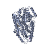 3rdeS C: citing same article ( S: Starting model for refinement |
|---|---|
| Similar structure data |


-Links
 PDBj
PDBj


- Assembly
| Deposited unit | 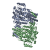
| ||||||||||||||||||
|---|---|---|---|---|---|---|---|---|---|---|---|---|---|---|---|---|---|---|---|
| 1 |
| ||||||||||||||||||
| Unit cell |
| ||||||||||||||||||
| Noncrystallographic symmetry (NCS) | NCS domain:
NCS domain segments: Component-ID: _ / Ens-ID: 1 / Beg auth comp-ID: VAL / Beg label comp-ID: VAL / End auth comp-ID: PRO / End label comp-ID: PRO / Refine code: _ / Auth seq-ID: 9 - 736 / Label seq-ID: 33 - 760
|
-Components
| #1: Protein | Mass: 86406.766 Da / Num. of mol.: 2 Source method: isolated from a genetically manipulated source Source: (gene. exp.) Gibberella zeae (strain PH-1 / ATCC MYA-4620 / FGSC 9075 / NRRL 31084) (fungus)Strain: PH-1 / ATCC MYA-4620 / FGSC 9075 / NRRL 31084 / Gene: FG02216.1, FGRAMPH1_01T05341 / Plasmid: pET-28b / Production host:  References: UniProt: I1REW2, Oxidoreductases; Acting on single donors with incorporation of molecular oxygen (oxygenases); With incorporation of two atoms of oxygen #2: Chemical |   Mass: 55.845 Da / Num. of mol.: 2 / Source method: obtained synthetically / Formula: Fe Mass: 55.845 Da / Num. of mol.: 2 / Source method: obtained synthetically / Formula: Fe#3: Water | ChemComp-HOH / |  Mass: 18.015 Da / Num. of mol.: 20 / Source method: isolated from a natural source / Formula: H2O Mass: 18.015 Da / Num. of mol.: 20 / Source method: isolated from a natural source / Formula: H2O |
|---|
-Experimental details
-Experiment
| Experiment | Method: X-RAY DIFFRACTION / Number of used crystals: 1 |
|---|
- Sample preparation
| Crystal | Density Matthews: 2.05 Å3/Da / Density % sol: 40.05 % |
|---|---|
| Crystal grow | Temperature: 295 K / Method: vapor diffusion, hanging drop / pH: 8.2 / Details: 22% PEG3350, 0.3 M ammonium acetate, 0.1 M Tris |
-Data collection
| Diffraction | Mean temperature: 100 K / Serial crystal experiment: N | |||||||||||||||
|---|---|---|---|---|---|---|---|---|---|---|---|---|---|---|---|---|
| Diffraction source | Source: SYNCHROTRON / Site: APS / Beamline: 24-ID-E / Wavelength: 0.97918 Å | |||||||||||||||
| Detector | Type: DECTRIS EIGER X 16M / Detector: PIXEL / Date: Dec 12, 2016 / Details: MIRRORS | |||||||||||||||
| Radiation | Monochromator: Cryogenically-cooled single crystal Si(220) side bounce Protocol: SINGLE WAVELENGTH / Monochromatic (M) / Laue (L): M / Scattering type: x-ray | |||||||||||||||
| Radiation wavelength | Wavelength: 0.97918 Å / Relative weight: 1 | |||||||||||||||
| Reflection twin |
| |||||||||||||||
| Reflection | Resolution: 2.79→83.59 Å / Num. obs: 33864 / % possible obs: 100 % / Redundancy: 3.8 % / Biso Wilson estimate: 41.7 Å2 / CC1/2: 0.992 / Rpim(I) all: 0.076 / Net I/σ(I): 8.6 | |||||||||||||||
| Reflection shell | Resolution: 2.79→2.94 Å / Redundancy: 3.6 % / Mean I/σ(I) obs: 1.9 / Num. unique obs: 4463 / CC1/2: 0.581 / Rpim(I) all: 0.428 / % possible all: 100 |
- Processing
| Software |
| ||||||||||||||||||||||||||||||||||||||||||||||||||||||||||||||||||||||||||||||||||||||||||||||||||||||||||||||||||||||||||||||||||||||||||||||||||||||||||||||||||||||||||||||||||||||
|---|---|---|---|---|---|---|---|---|---|---|---|---|---|---|---|---|---|---|---|---|---|---|---|---|---|---|---|---|---|---|---|---|---|---|---|---|---|---|---|---|---|---|---|---|---|---|---|---|---|---|---|---|---|---|---|---|---|---|---|---|---|---|---|---|---|---|---|---|---|---|---|---|---|---|---|---|---|---|---|---|---|---|---|---|---|---|---|---|---|---|---|---|---|---|---|---|---|---|---|---|---|---|---|---|---|---|---|---|---|---|---|---|---|---|---|---|---|---|---|---|---|---|---|---|---|---|---|---|---|---|---|---|---|---|---|---|---|---|---|---|---|---|---|---|---|---|---|---|---|---|---|---|---|---|---|---|---|---|---|---|---|---|---|---|---|---|---|---|---|---|---|---|---|---|---|---|---|---|---|---|---|---|---|
| Refinement | Method to determine structure: MOLECULAR REPLACEMENT Starting model: PDB entry 3RDE Resolution: 2.79→64.08 Å / Cor.coef. Fo:Fc: 0.934 / Cor.coef. Fo:Fc free: 0.877 / SU B: 24.908 / SU ML: 0.241 / Cross valid method: THROUGHOUT / ESU R Free: 0.081 / Details: HYDROGENS HAVE BEEN ADDED IN THE RIDING POSITIONS
| ||||||||||||||||||||||||||||||||||||||||||||||||||||||||||||||||||||||||||||||||||||||||||||||||||||||||||||||||||||||||||||||||||||||||||||||||||||||||||||||||||||||||||||||||||||||
| Solvent computation | Ion probe radii: 0.8 Å / Shrinkage radii: 0.8 Å / VDW probe radii: 1.2 Å | ||||||||||||||||||||||||||||||||||||||||||||||||||||||||||||||||||||||||||||||||||||||||||||||||||||||||||||||||||||||||||||||||||||||||||||||||||||||||||||||||||||||||||||||||||||||
| Displacement parameters | Biso mean: 47.232 Å2
| ||||||||||||||||||||||||||||||||||||||||||||||||||||||||||||||||||||||||||||||||||||||||||||||||||||||||||||||||||||||||||||||||||||||||||||||||||||||||||||||||||||||||||||||||||||||
| Refinement step | Cycle: 1 / Resolution: 2.79→64.08 Å
| ||||||||||||||||||||||||||||||||||||||||||||||||||||||||||||||||||||||||||||||||||||||||||||||||||||||||||||||||||||||||||||||||||||||||||||||||||||||||||||||||||||||||||||||||||||||
| Refine LS restraints |
|