 Movie
Movie Controller

[English] 日本語
 Yorodumi
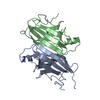
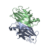
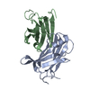
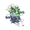
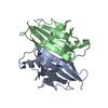
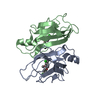
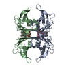
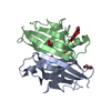
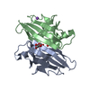
- PDB-1e3f: Structure of human transthyretin complexed with bromophenols: a n... -
+ Open data

- Basic information
| Entry | Database: PDB / ID: 1e3f | ||||||
|---|---|---|---|---|---|---|---|
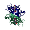
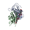
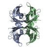
| Title | Structure of human transthyretin complexed with bromophenols: a new mode of binding | ||||||
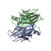
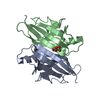
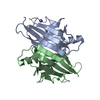
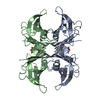
 Components Components | TRANSTHYRETIN | ||||||
 Keywords Keywords | TRANSPORT(THYROXINE) / ENVIRONMENTAL POLLUTANTS / BROMOPHENOLS | ||||||
| Function / homology |  Function and homology information Function and homology informationDefective visual phototransduction due to STRA6 loss of function / negative regulation of glomerular filtration / The canonical retinoid cycle in rods (twilight vision) / purine nucleobase metabolic process / hormone binding / Non-integrin membrane-ECM interactions / phototransduction, visible light / molecular sequestering activity / retinoid metabolic process / Retinoid metabolism and transport ...Defective visual phototransduction due to STRA6 loss of function / negative regulation of glomerular filtration / The canonical retinoid cycle in rods (twilight vision) / purine nucleobase metabolic process / hormone binding / Non-integrin membrane-ECM interactions / phototransduction, visible light / molecular sequestering activity / retinoid metabolic process / Retinoid metabolism and transport / hormone activity / azurophil granule lumen / Amyloid fiber formation / Neutrophil degranulation / protein-containing complex binding / protein-containing complex / : / extracellular exosome / extracellular region / identical protein binding Similarity search - Function | ||||||
| Biological species |  HOMO SAPIENS (human) HOMO SAPIENS (human) | ||||||
| Method |  X-RAY DIFFRACTION / MOLECULAR REPLACEMENT / Resolution: 1.9 Å X-RAY DIFFRACTION / MOLECULAR REPLACEMENT / Resolution: 1.9 Å | ||||||
 Authors Authors | Ghosh, M. / Meerts, I.A.T.M. / Cook, A. / Bergman, A. / Brouwer, A. / Johnson, L.N. | ||||||
 Citation Citation | Journal: Acta Crystallogr.,Sect.D / Year: 2000 Title: Structure of Human Transthyretin Complexed with Bromophenols : A New Mode of Binding Authors: Ghosh, M. / Meerts, I.A.T.M. / Cook, A. / Bergman, A. / Brouwer, A. / Johnson, L.N. #1: Journal: The Design of Drugs to Macromolecular Targets / Year: 1992Title: Multiple Modes of Binding of Thyroid Hormones and Other Iodothyronines to Human Plasma Transthyretin Authors: De La Paz, P. / Burridge, J.M. / Oatley, S.J. / Blake, C.C.F. #2: Journal: J.Mol.Biol. / Year: 1978Title: Structure of Prealbumin.Secondary,Tertiary and Quaternary Interactions Determined by Fourier Refinemrnt and Thyroxine Binding Authors: Blake, C.C.F. / Geisow, M.J. / Oatley, S.J. / Rerat, C. / Rerat, B. #3: Journal: Nature / Year: 1977 Title: Protein-DNA and Protein-Hormone Interactions in Prealbumin : A Model of the Thyroid Hormone Nuclear Receptor Authors: Blake, C.C.F. / Oatley, S.J. | ||||||
| History |
|
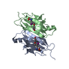
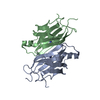
- Structure visualization
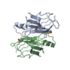
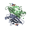
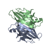
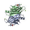
| Structure viewer | Molecule: MolmilJmol/JSmol |
|---|
- Downloads & links
-Download
| PDBx/mmCIF format | 1e3f.cif.gz | 56.4 KB | Display | PDBx/mmCIF format |
|---|---|---|---|---|
| PDB format | pdb1e3f.ent.gz | 40.9 KB | Display | PDB format |
| PDBx/mmJSON format | 1e3f.json.gz | Tree view | PDBx/mmJSON format | |
| Others |  Other downloads Other downloads |
-Validation report
| Arichive directory | https://data.pdbj.org/pub/pdb/validation_reports/e3/1e3fftp://data.pdbj.org/pub/pdb/validation_reports/e3/1e3f | HTTPS FTP |
|---|
-Related structure data
| Related structure data | 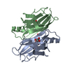 1e4hC 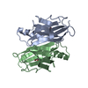 1e5aC  1ttaS S: Starting model for refinement C: citing same article ( |
|---|---|
| Similar structure data |


-Links
 PDBj
PDBj


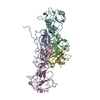
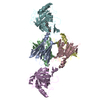
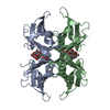
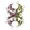
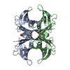
- Assembly
| Deposited unit | 
| |||||||||
|---|---|---|---|---|---|---|---|---|---|---|
| 1 | 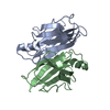
| |||||||||
| Unit cell |
| |||||||||
| Components on special symmetry positions |
| |||||||||
| Noncrystallographic symmetry (NCS) | NCS oper: (Code: given Matrix: (-0.9898, -0.1422, 0.0087), Vector: |
-Components
| #1: Protein | Mass: 13777.360 Da / Num. of mol.: 2 / Source method: isolated from a natural source / Source: (natural) HOMO SAPIENS (human) / Organ: PLASMA / References: UniProt: P02766#2: Water | ChemComp-HOH / |  Mass: 18.015 Da / Num. of mol.: 78 / Source method: isolated from a natural source / Formula: H2O Mass: 18.015 Da / Num. of mol.: 78 / Source method: isolated from a natural source / Formula: H2O |
|---|
-Experimental details
-Experiment
| Experiment | Method: X-RAY DIFFRACTION / Number of used crystals: 1 |
|---|
- Sample preparation
| Crystal | Density Matthews: 2.2 Å3/Da / Density % sol: 43 % | ||||||||||||||||||||||||||||||||||||||||||
|---|---|---|---|---|---|---|---|---|---|---|---|---|---|---|---|---|---|---|---|---|---|---|---|---|---|---|---|---|---|---|---|---|---|---|---|---|---|---|---|---|---|---|---|
| Crystal grow | pH: 5.5 / Details: pH 5.50 | ||||||||||||||||||||||||||||||||||||||||||
| Crystal grow | *PLUS Temperature: 295 K / pH: 8 / Method: vapor diffusion, hanging drop | ||||||||||||||||||||||||||||||||||||||||||
| Components of the solutions | *PLUS
|
-Data collection
| Diffraction | Mean temperature: 100 K |
|---|---|
| Diffraction source | Source: ROTATING ANODE / Type: RIGAKU RUH2R / Wavelength: 1.5418 |
| Detector | Type: MARRESEARCH / Detector: IMAGE PLATE / Date: Feb 15, 1997 / Details: YALE MIRRORS |
| Radiation | Protocol: SINGLE WAVELENGTH / Monochromatic (M) / Laue (L): M / Scattering type: x-ray |
| Radiation wavelength | Wavelength: 1.5418 Å / Relative weight: 1 |
| Reflection | Resolution: 1.9→20 Å / Num. obs: 16505 / % possible obs: 85.7 % / Redundancy: 3.2 % / Biso Wilson estimate: 28.3 Å2 / Rmerge(I) obs: 0.039 / Rsym value: 0.039 / Net I/σ(I): 22.1 |
| Reflection shell | Resolution: 1.9→1.99 Å / Redundancy: 3 % / Rmerge(I) obs: 0.228 / Mean I/σ(I) obs: 3.6 / Rsym value: 0.228 / % possible all: 88.7 |
| Reflection | *PLUS Num. measured all: 215400 |
| Reflection shell | *PLUS % possible obs: 88.7 % / Num. unique obs: 2102 |
- Processing
| Software |
| ||||||||||||||||||||||||||||||||||||||||||||||||||||||||||||
|---|---|---|---|---|---|---|---|---|---|---|---|---|---|---|---|---|---|---|---|---|---|---|---|---|---|---|---|---|---|---|---|---|---|---|---|---|---|---|---|---|---|---|---|---|---|---|---|---|---|---|---|---|---|---|---|---|---|---|---|---|---|
| Refinement | Method to determine structure: MOLECULAR REPLACEMENT Starting model: 1TTA Resolution: 1.9→20 Å / Data cutoff high absF: 0 / Data cutoff low absF: 0 / Cross valid method: THROUGHOUT / σ(F): 0 Details: RESIDUES 1-9 OF BOTH CHAINS, AS WELL AS 126-127 OF CHAIN A AND 125-127 OF CHAIN B ARE ILL-DEFINED IN THE ELECTRON DENSITY MAPS AND HAVE BEEN OMITTED. THERE ARE SEVERAL OTHER RESIDUES WHERE ...Details: RESIDUES 1-9 OF BOTH CHAINS, AS WELL AS 126-127 OF CHAIN A AND 125-127 OF CHAIN B ARE ILL-DEFINED IN THE ELECTRON DENSITY MAPS AND HAVE BEEN OMITTED. THERE ARE SEVERAL OTHER RESIDUES WHERE THE MAIN CHAIN WAS VISIBLE BUT THE SIDE CHAIN DENSITIES WERE POOR. THESE RESIDUES HAVE BEEN REPLACED BY ALANINES. THIS COORDINATE SET COMPRISES TWO CHAINS REPRESENTING TWO CHEMICALLY EQUIVALENT, BUT CRYSTALLOGRAPHICALLY DISTINCT, ENTITIES. THE OTHER HALF OF THE COMPLETE TETRAMER MAY BE GENERATED FROM THIS DIMER BY THE APPLICATION OF THE CRYSTALLOGRAPHIC DIAD PARALLEL TO Z THROUGH THE ORIGIN OF THIS COORDINATE SYSTEM, I. E. XPRIME=-X, YPRIME= -Y, ZPRIME= Z. THERE ARE WATER MOLECULES LOCATED ON THE TWO- FOLD SYMMETRY AXIS (W47, W87) WHICH ARE ASSIGNED 50% OCCUPANCY.
| ||||||||||||||||||||||||||||||||||||||||||||||||||||||||||||
| Refinement step | Cycle: LAST / Resolution: 1.9→20 Å
| ||||||||||||||||||||||||||||||||||||||||||||||||||||||||||||
| Refine LS restraints |
| ||||||||||||||||||||||||||||||||||||||||||||||||||||||||||||
| LS refinement shell | Resolution: 1.9→2.05 Å / Total num. of bins used: 5
| ||||||||||||||||||||||||||||||||||||||||||||||||||||||||||||
| Xplor file | Serial no: 1 / Param file: PARHCSDX.PRO / Topol file: TOPHCSDX.PRO | ||||||||||||||||||||||||||||||||||||||||||||||||||||||||||||
| Software | *PLUS Name: X-PLOR / Version: 3.851 / Classification: refinement | ||||||||||||||||||||||||||||||||||||||||||||||||||||||||||||
| Refine LS restraints | *PLUS
|
