 Movie
Movie Controller


+ Open data

- Basic information
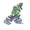
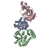
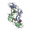
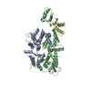
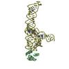
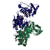
| Entry | Database: PDB / ID: 1rlb | ||||||
|---|---|---|---|---|---|---|---|
| Title | RETINOL BINDING PROTEIN COMPLEXED WITH TRANSTHYRETIN | ||||||
 Components Components |
| ||||||
 Keywords Keywords | COMPLEX (PROTEIN/PROTEIN) / COMPLEX (PROTEIN-PROTEIN) / COMPLEX (PROTEIN-PROTEIN) complex | ||||||
| Function / homology |  Function and homology information Function and homology informationRetinoid metabolism disease events / urinary bladder development / vitamin A import into cell / embryonic retina morphogenesis in camera-type eye / retinol transport / female genitalia morphogenesis / retinol transmembrane transporter activity / maintenance of gastrointestinal epithelium / embryonic organ morphogenesis / negative regulation of cardiac muscle cell proliferation ...Retinoid metabolism disease events / urinary bladder development / vitamin A import into cell / embryonic retina morphogenesis in camera-type eye / retinol transport / female genitalia morphogenesis / retinol transmembrane transporter activity / maintenance of gastrointestinal epithelium / embryonic organ morphogenesis / negative regulation of cardiac muscle cell proliferation / embryonic skeletal system development / detection of light stimulus involved in visual perception / retinal metabolic process / eye development / retinal binding / heart trabecula formation / retinol metabolic process / molecular carrier activity / retinol binding / cardiac muscle tissue development / Defective visual phototransduction due to STRA6 loss of function / negative regulation of glomerular filtration / positive regulation of immunoglobulin production / response to muscle activity / The canonical retinoid cycle in rods (twilight vision) / uterus development / vagina development / purine nucleobase metabolic process / hormone binding / Non-integrin membrane-ECM interactions / phototransduction, visible light / molecular sequestering activity / response to retinoic acid / retinoid metabolic process / Retinoid metabolism and transport / lung development / gluconeogenesis / hormone activity / response to insulin / male gonad development / positive regulation of insulin secretion / azurophil granule lumen / glucose homeostasis / heart development / spermatogenesis / response to ethanol / response to xenobiotic stimulus / Amyloid fiber formation / Neutrophil degranulation / protein-containing complex binding / protein-containing complex / : / extracellular exosome / extracellular region / identical protein binding Similarity search - Function | ||||||
| Biological species |  Homo sapiens (human) Homo sapiens (human) | ||||||
| Method |  X-RAY DIFFRACTION / Resolution: 3.1 Å X-RAY DIFFRACTION / Resolution: 3.1 Å | ||||||
 Authors Authors | Monaco, H.L. / Rizzi, M. / Coda, A. | ||||||
 Citation Citation | Journal: Science / Year: 1995 Title: Structure of a complex of two plasma proteins: transthyretin and retinol-binding protein. Authors: Monaco, H.L. / Rizzi, M. / Coda, A. #1: Journal: J.Mol.Biol. / Year: 1994Title: Crystallization of the Macromolecular Complex Transthyretin Retinol Binding Protein Authors: Monaco, H.L. / Mancia, F. / Rizzi, M. / Coda, A. | ||||||
| History |
|
- Structure visualization
| Structure viewer | Molecule: MolmilJmol/JSmol |
|---|
- Downloads & links
-Download
| PDBx/mmCIF format | 1rlb.cif.gz | 160.3 KB | Display | PDBx/mmCIF format |
|---|---|---|---|---|
| PDB format | pdb1rlb.ent.gz | 128.3 KB | Display | PDB format |
| PDBx/mmJSON format | 1rlb.json.gz | Tree view | PDBx/mmJSON format | |
| Others |  Other downloads Other downloads |
-Validation report
| Arichive directory | https://data.pdbj.org/pub/pdb/validation_reports/rl/1rlbftp://data.pdbj.org/pub/pdb/validation_reports/rl/1rlb | HTTPS FTP |
|---|
-Related structure data
| Similar structure data |
|---|


-Links
 PDBj
PDBj


- Assembly
| Deposited unit | 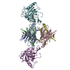
| ||||||||
|---|---|---|---|---|---|---|---|---|---|
| 1 |
| ||||||||
| Unit cell |
|
-Components
| #1: Protein | Mass: 13776.376 Da / Num. of mol.: 4 / Source method: isolated from a natural source / Source: (natural) Homo sapiens (human) / Organ: PLASMA / References: UniProt: P02766#2: Protein | Mass: 20079.545 Da / Num. of mol.: 2 / Source method: isolated from a natural source / Source: (natural) #3: Chemical |   Mass: 300.435 Da / Num. of mol.: 2 / Source method: obtained synthetically / Formula: C20H28O2 Mass: 300.435 Da / Num. of mol.: 2 / Source method: obtained synthetically / Formula: C20H28O2Has protein modification | Y | |
|---|
-Experimental details
-Experiment
| Experiment | Method: X-RAY DIFFRACTION / Number of used crystals: 1 |
|---|
- Sample preparation
| Crystal | Density Matthews: 2.65 Å3/Da / Density % sol: 53.5 % | |||||||||||||||||||||||||
|---|---|---|---|---|---|---|---|---|---|---|---|---|---|---|---|---|---|---|---|---|---|---|---|---|---|---|
| Crystal | *PLUS Density % sol: 53.5 % | |||||||||||||||||||||||||
| Crystal grow | *PLUS Temperature: 4 ℃ / pH: 5.5 / Method: microdialysis | |||||||||||||||||||||||||
| Components of the solutions | *PLUS
|
-Data collection
| Diffraction source | Wavelength: 1.5418 Å |
|---|---|
| Detector | Type: RIGAKU / Detector: IMAGE PLATE |
| Radiation | Monochromatic (M) / Laue (L): M / Scattering type: x-ray |
| Radiation wavelength | Wavelength: 1.5418 Å / Relative weight: 1 |
| Reflection | Num. obs: 17465 / % possible obs: 94.8 % / Observed criterion σ(I): 0 / Redundancy: 2.7 % / Rmerge(I) obs: 0.09 |
| Reflection | *PLUS Highest resolution: 3.1 Å / Lowest resolution: 6 Å / Num. measured all: 47491 / Rmerge(I) obs: 0.09 |
| Reflection shell | *PLUS Highest resolution: 3.1 Å / Lowest resolution: 3.3 Å / % possible obs: 83.2 % / Num. unique obs: 2359 |
- Processing
| Software |
| ||||||||||||||||||||||||||||||||||||||||||||||||||||||||||||
|---|---|---|---|---|---|---|---|---|---|---|---|---|---|---|---|---|---|---|---|---|---|---|---|---|---|---|---|---|---|---|---|---|---|---|---|---|---|---|---|---|---|---|---|---|---|---|---|---|---|---|---|---|---|---|---|---|---|---|---|---|---|
| Refinement | Resolution: 3.1→6 Å / σ(F): 0 Details: THERE IS CLOSE CONTACT BETWEEN RESIDUE GLU D 66 AND A SYMMETRY-RELATED COPY OF ITSELF.
| ||||||||||||||||||||||||||||||||||||||||||||||||||||||||||||
| Refinement step | Cycle: LAST / Resolution: 3.1→6 Å
| ||||||||||||||||||||||||||||||||||||||||||||||||||||||||||||
| Refine LS restraints |
| ||||||||||||||||||||||||||||||||||||||||||||||||||||||||||||
| Software | *PLUS Name: X-PLOR / Classification: refinement | ||||||||||||||||||||||||||||||||||||||||||||||||||||||||||||
| Refinement | *PLUS Rfactor Rwork: 0.201 | ||||||||||||||||||||||||||||||||||||||||||||||||||||||||||||
| Solvent computation | *PLUS | ||||||||||||||||||||||||||||||||||||||||||||||||||||||||||||
| Displacement parameters | *PLUS | ||||||||||||||||||||||||||||||||||||||||||||||||||||||||||||
| Refine LS restraints | *PLUS Type: x_dihedral_angle_deg / Dev ideal: 26.7 |
