 Movie
Movie Controller

[English] 日本語
 Yorodumi
- PDB-2pab: STRUCTURE OF PREALBUMIN, SECONDARY, TERTIARY AND QUATERNARY INTER... -
+ Open data

- Basic information
| Entry | Database: PDB / ID: 2pab | ||||||||||||
|---|---|---|---|---|---|---|---|---|---|---|---|---|---|
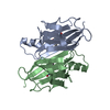
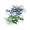
| Title | STRUCTURE OF PREALBUMIN, SECONDARY, TERTIARY AND QUATERNARY INTERACTIONS DETERMINED BY FOURIER REFINEMENT AT 1.8 ANGSTROMS | ||||||||||||
 Components Components | TRANSTHYRETIN PRECURSOR | ||||||||||||
 Keywords Keywords | TRANSPORT (THYROXINE / RETINOL) IN SERUM | ||||||||||||
| Function / homology |  Function and homology information Function and homology informationDefective visual phototransduction due to STRA6 loss of function / negative regulation of glomerular filtration / The canonical retinoid cycle in rods (twilight vision) / purine nucleobase metabolic process / hormone binding / Non-integrin membrane-ECM interactions / phototransduction, visible light / molecular sequestering activity / retinoid metabolic process / Retinoid metabolism and transport ...Defective visual phototransduction due to STRA6 loss of function / negative regulation of glomerular filtration / The canonical retinoid cycle in rods (twilight vision) / purine nucleobase metabolic process / hormone binding / Non-integrin membrane-ECM interactions / phototransduction, visible light / molecular sequestering activity / retinoid metabolic process / Retinoid metabolism and transport / hormone activity / azurophil granule lumen / Amyloid fiber formation / Neutrophil degranulation / protein-containing complex binding / protein-containing complex / : / extracellular exosome / extracellular region / identical protein binding Similarity search - Function | ||||||||||||
| Biological species |  Homo sapiens (human) Homo sapiens (human) | ||||||||||||
| Method |  X-RAY DIFFRACTION / Resolution: 1.8 Å X-RAY DIFFRACTION / Resolution: 1.8 Å | ||||||||||||
 Authors Authors | Oatley, S.J. / Blake, C.C.F. | ||||||||||||
 Citation Citation | Journal: J.Mol.Biol. / Year: 1978 Title: Structure of prealbumin: secondary, tertiary and quaternary interactions determined by Fourier refinement at 1.8 A. Authors: Blake, C.C. / Geisow, M.J. / Oatley, S.J. / Rerat, B. / Rerat, C. #1: Journal: Nature / Year: 1977Title: Protein-DNA and Protein-Hormone Interactions in Prealbumin,A Model of the Thyroid Hormone Nuclear Receptor (Query) Authors: Blake, C.C.F. / Oatley, S.J. #2: Journal: J.Mol.Biol. / Year: 1974Title: Structure of Human Plasma Prealbumin at 2.5 Angstroms Resolution,A Preliminary Report on the Polypeptide Chain Conformation,Quaternary Structure and Thyroxine Binding Authors: Blake, C.C.F. / Geisow, M.J. / Swan, I.D.A. / Rerat, C. / Rerat, B. #3: Journal: J.Mol.Biol. / Year: 1971Title: An X-Ray Study of the Subunit Structure of Prealbumin Authors: Blake, C.C.F. / Swan, I.D.A. / Rerat, C. / Berthou, J. / Laurent, A. / Rerat, B. | ||||||||||||
| History |
| ||||||||||||
| Remark 700 | SHEET THE FIRST STRAND OF THE *EXTERNAL* SHEET IN EACH SUBUNIT IS DISCONTINUOUS. TO ACCOMODATE ...SHEET THE FIRST STRAND OF THE *EXTERNAL* SHEET IN EACH SUBUNIT IS DISCONTINUOUS. TO ACCOMODATE THESE DISCONTINUITIES EACH *EXTERNAL* SHEET IS REPRESENTED HERE TWICE, WITH A DIFFERENT STRAND 1 IN EACH CASE. STRANDS 2,3,4 OF *X1A* ARE IDENTICAL TO STRANDS 2,3,4 OF *X2A*. SIMILARLY STRANDS 2,3,4 OF *X1B* ARE IDENTICAL TO STRANDS 2,3,4 OF *X2B*. DESPITE THIS PARTIAL REDUNDANCY OF REPRESENTATION THERE IS NO IMPLICATION THAT EACH SUBUNIT CONTAINS MORE THAN ONE *EXTERNAL* SHEET SUBSTRUCTURE. |
- Structure visualization
| Structure viewer | Molecule: MolmilJmol/JSmol |
|---|
- Downloads & links
-Download
| PDBx/mmCIF format | 2pab.cif.gz | 51.8 KB | Display | PDBx/mmCIF format |
|---|---|---|---|---|
| PDB format | pdb2pab.ent.gz | 36.1 KB | Display | PDB format |
| PDBx/mmJSON format | 2pab.json.gz | Tree view | PDBx/mmJSON format | |
| Others |  Other downloads Other downloads |
-Validation report
| Arichive directory | https://data.pdbj.org/pub/pdb/validation_reports/pa/2pabftp://data.pdbj.org/pub/pdb/validation_reports/pa/2pab | HTTPS FTP |
|---|
-Related structure data
| Similar structure data |
|---|


-Links
 PDBj
PDBj


- Assembly
| Deposited unit | 
| ||||||||
|---|---|---|---|---|---|---|---|---|---|
| 1 |
| ||||||||
| 2 | 
| ||||||||
| Unit cell |
| ||||||||
| Noncrystallographic symmetry (NCS) | NCS oper: (Code: given / Matrix: (-0.98771, 0.156297),| Details | THIS COORDINATE SET COMPRISES TWO CHAINS REPRESENTING TWO CHEMICALLY EQUIVALENT, BUT CRYSTALLOGRAPHICALLY DISTINCT, ENTITIES. THE OTHER HALF OF THE COMPLETE TETRAMER MAY BE GENERATED FROM THIS DIMER BY THE APPLICATION OF THE CRYSTALLOGRAPHIC DIAD PARALLEL TO Z THROUGH THE ORIGIN OF THIS COORDINATE SYSTEM, I. E. XPRIME=-X, YPRIME=-Y, ZPRIME=Z. | |
-Components
| #1: Protein | Mass: 13776.376 Da / Num. of mol.: 2 Source method: isolated from a genetically manipulated source Source: (gene. exp.) Homo sapiens (human) / Production host:  |
|---|
-Experimental details
-Experiment
| Experiment | Method: X-RAY DIFFRACTION |
|---|
- Sample preparation
| Crystal | Density Matthews: 2.23 Å3/Da / Density % sol: 44.84 % |
|---|---|
| Crystal grow | *PLUS Method: other / Details: Blake, C.C.F., (1974) J. Mol. Biol., 88, 1. |
-Data collection
| Radiation | Scattering type: x-ray |
|---|---|
| Radiation wavelength | Relative weight: 1 |
| Reflection | Resolution: 1.8→15 Å |
- Processing
| Refinement | Resolution: 1.8→8 Å / Rfactor Rwork: 0.29 / Rfactor obs: 0.29 Details: THE AXES OF THE COORDINATE SYSTEM USED HERE ARE ALIGNED WITH THE CRYSTALLOGRAPHIC AXES BUT THE ORIGIN IS DISPLACED BY A/2, B/2, C/4 AS GIVEN IN THE SCALE RECORDS BELOW. | ||||||||||||
|---|---|---|---|---|---|---|---|---|---|---|---|---|---|
| Refinement step | Cycle: LAST / Resolution: 1.8→8 Å
| ||||||||||||
| Refinement | *PLUS Rfactor obs: 0.289 | ||||||||||||
| Solvent computation | *PLUS | ||||||||||||
| Displacement parameters | *PLUS |
