 Movie
Movie Controller

[English] 日本語
 Yorodumi
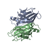
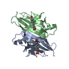
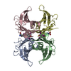
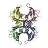
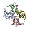
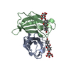
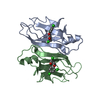
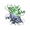
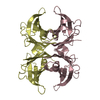
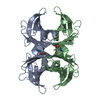
- PDB-1thc: CRYSTAL STRUCTURE DETERMINATION AT 2.3A OF HUMAN TRANSTHYRETIN-3'... -
+ Open data

- Basic information
| Entry | Database: PDB / ID: 1thc | ||||||
|---|---|---|---|---|---|---|---|
| Title | CRYSTAL STRUCTURE DETERMINATION AT 2.3A OF HUMAN TRANSTHYRETIN-3',5'-DIBROMO-2',4,4',6-TETRA-HYDROXYAURONE COMPLEX | ||||||
 Components Components | TRANSTHYRETIN | ||||||
 Keywords Keywords | TRANSPORT (THYROXINE / RETINOL) IN SERUM | ||||||
| Function / homology |  Function and homology information Function and homology informationDefective visual phototransduction due to STRA6 loss of function / negative regulation of glomerular filtration / The canonical retinoid cycle in rods (twilight vision) / purine nucleobase metabolic process / hormone binding / Non-integrin membrane-ECM interactions / phototransduction, visible light / molecular sequestering activity / retinoid metabolic process / Retinoid metabolism and transport ...Defective visual phototransduction due to STRA6 loss of function / negative regulation of glomerular filtration / The canonical retinoid cycle in rods (twilight vision) / purine nucleobase metabolic process / hormone binding / Non-integrin membrane-ECM interactions / phototransduction, visible light / molecular sequestering activity / retinoid metabolic process / Retinoid metabolism and transport / hormone activity / azurophil granule lumen / Amyloid fiber formation / Neutrophil degranulation / protein-containing complex binding / protein-containing complex / : / extracellular exosome / extracellular region / identical protein binding Similarity search - Function | ||||||
| Biological species |  Homo sapiens (human) Homo sapiens (human) | ||||||
| Method |  X-RAY DIFFRACTION / Resolution: 2.3 Å X-RAY DIFFRACTION / Resolution: 2.3 Å | ||||||
 Authors Authors | Ciszak, E. / Cody, V. / Luft, J.R. | ||||||
 Citation Citation | Journal: Proc.Natl.Acad.Sci.USA / Year: 1992 Title: Crystal structure determination at 2.3-A resolution of human transthyretin-3',5'-dibromo-2',4,4',6-tetrahydroxyaurone complex. Authors: Ciszak, E. / Cody, V. / Luft, J.R. #1: Journal: J.Biol.Chem. / Year: 1992Title: Mechanism of Molecular Recognition. Structural Aspects of 3,3'-Diiodo-L-Thyronine Binding to Human Serum Transthyretin Authors: Wojtczak, A. / Luft, J. / Cody, V. #2: Journal: Nature / Year: 1977Title: Protein-DNA and Protein-Hormone Interactions in Prealbumin, a Model of the Thyroid Hormone Nuclear Receptor (Query) Authors: Blake, C.C.F. / Oatley, S.J. #3: Journal: J.Mol.Biol. / Year: 1974Title: Structure of Human Plasma Prealbumin at 2.5 Angstroms Resolution,A Preliminary Report on the Polypeptide Chain Conformation,Quaternary Structure and Thyroxine Binding Authors: Blake, C.C.F. / Geisow, M.J. / Swan, I.D.A. / Rerat, C. / Rerat, B. #4: Journal: J.Mol.Biol. / Year: 1971Title: An X-Ray Study of the Subunit Structure of Prealbumin Authors: Blake, C.C.F. / Swan, I.D.A. / Rerat, C. / Berthou, J. / Laurent, A. / Rerat, B. | ||||||
| History |
| ||||||
| Remark 700 | SHEET THE FIRST STRAND OF THE *EXTERNAL* SHEET IN EACH SUBUNIT IS DISCONTINUOUS. TO ACCOMMODATE ...SHEET THE FIRST STRAND OF THE *EXTERNAL* SHEET IN EACH SUBUNIT IS DISCONTINUOUS. TO ACCOMMODATE THESE DISCONTINUITIES EACH *EXTERNAL* SHEET IS REPRESENTED HERE TWICE, WITH A DIFFERENT STRAND 1 IN EACH CASE. STRANDS 2,3,4 OF *X1A* ARE IDENTICAL TO STRANDS 2,3,4 OF *X2A*. SIMILARLY STRANDS 2,3,4 OF *X1B* ARE IDENTICAL TO STRANDS 2,3,4 OF *X2B*. THIS DESCRIPTION IS CONSISTENT WITH THAT USED BY BLAKE AND COWORKERS FOR THE NATIVE TRANSTHYRETIN (PROTEIN DATA BANK ENTRY 2PAB). |
- Structure visualization
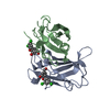
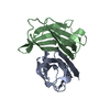
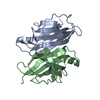
| Structure viewer | Molecule: MolmilJmol/JSmol |
|---|
- Downloads & links
-Download
| PDBx/mmCIF format | 1thc.cif.gz | 63.5 KB | Display | PDBx/mmCIF format |
|---|---|---|---|---|
| PDB format | pdb1thc.ent.gz | 46.4 KB | Display | PDB format |
| PDBx/mmJSON format | 1thc.json.gz | Tree view | PDBx/mmJSON format | |
| Others |  Other downloads Other downloads |
-Validation report
| Arichive directory | https://data.pdbj.org/pub/pdb/validation_reports/th/1thcftp://data.pdbj.org/pub/pdb/validation_reports/th/1thc | HTTPS FTP |
|---|
-Related structure data
| Similar structure data |
|---|


-Links
 PDBj
PDBj


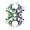
- Assembly
| Deposited unit | 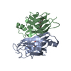
| ||||||||||||||||||||||||||||||||||||
|---|---|---|---|---|---|---|---|---|---|---|---|---|---|---|---|---|---|---|---|---|---|---|---|---|---|---|---|---|---|---|---|---|---|---|---|---|---|
| 1 | 
| ||||||||||||||||||||||||||||||||||||
| Unit cell |
| ||||||||||||||||||||||||||||||||||||
| Components on special symmetry positions |
|
-Components
| #1: Protein | Mass: 13776.376 Da / Num. of mol.: 2 Source method: isolated from a genetically manipulated source Source: (gene. exp.) Homo sapiens (human) / References: UniProt: P02766#2: Chemical | ChemComp-FL9 / 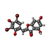  Mass: 444.028 Da / Num. of mol.: 4 / Source method: obtained synthetically / Formula: C15H8Br2O6 Mass: 444.028 Da / Num. of mol.: 4 / Source method: obtained synthetically / Formula: C15H8Br2O6#3: Water | ChemComp-HOH / |  Mass: 18.015 Da / Num. of mol.: 95 / Source method: isolated from a natural source / Formula: H2O Mass: 18.015 Da / Num. of mol.: 95 / Source method: isolated from a natural source / Formula: H2ONonpolymer details | THE STRUCTURE CONTAINS THE 3',5'-DIBROMO-2',4,4',6-TETRAHYDROXY AURONE MOLECULES THAT BIND IN TWO ...THE STRUCTURE CONTAINS THE 3',5'-DIBROMO-2',4,4',6-TETRAHYDRO | |
|---|
-Experimental details
-Experiment
| Experiment | Method: X-RAY DIFFRACTION |
|---|
- Sample preparation
| Crystal | Density Matthews: 2.22 Å3/Da / Density % sol: 44.65 % | |||||||||||||||
|---|---|---|---|---|---|---|---|---|---|---|---|---|---|---|---|---|
| Crystal grow | *PLUS pH: 6.9 / Method: batch method | |||||||||||||||
| Components of the solutions | *PLUS
|
-Data collection
| Reflection | *PLUS Num. obs: 11830 / Num. measured all: 18899 / Rmerge(I) obs: 0.05 |
|---|
- Processing
| Software | Name: PROLSQ / Classification: refinement | ||||||||||||||||||||||||||||
|---|---|---|---|---|---|---|---|---|---|---|---|---|---|---|---|---|---|---|---|---|---|---|---|---|---|---|---|---|---|
| Refinement | Rfactor obs: 0.179 / Highest resolution: 2.3 Å | ||||||||||||||||||||||||||||
| Refinement step | Cycle: LAST / Highest resolution: 2.3 Å
| ||||||||||||||||||||||||||||
| Refine LS restraints |
| ||||||||||||||||||||||||||||
| Refinement | *PLUS Highest resolution: 2.3 Å / Lowest resolution: 10 Å / Num. reflection obs: 8442 / σ(F): 2 / Rfactor all: 0.179 | ||||||||||||||||||||||||||||
| Solvent computation | *PLUS | ||||||||||||||||||||||||||||
| Displacement parameters | *PLUS | ||||||||||||||||||||||||||||
| Refine LS restraints | *PLUS
|
