[English] 日本語
 Yorodumi
- PDB-1z7j: Human transthyretin (also called prealbumin) complex with 3, 3',5... -
+ Open data
- Basic information
| Entry | Database: PDB / ID: 1z7j | |||||||||
|---|---|---|---|---|---|---|---|---|---|---|
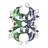
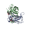
| Title | Human transthyretin (also called prealbumin) complex with 3, 3',5,5'-tetraiodothyroacetic acid (t4ac) | |||||||||
 Components Components | Transthyretin | |||||||||
 Keywords Keywords | TRANSPORT PROTEIN / ALBUMIN / TRANSPORT / RETINOL-BINDING / VITAMIN A / AMYLOID / THYROID HORMONE / LIVER / PLASMA / CEREBROSPINAL FLUID / POLYNEUROPATHY / DISEASE MUTATION / TETRAIODOTHYROACETIC ACID / T4AC / PREALBUMIN | |||||||||
| Function / homology |  Function and homology information Function and homology informationDefective visual phototransduction due to STRA6 loss of function / negative regulation of glomerular filtration / The canonical retinoid cycle in rods (twilight vision) / purine nucleobase metabolic process / hormone binding / Non-integrin membrane-ECM interactions / molecular sequestering activity / phototransduction, visible light / retinoid metabolic process / Retinoid metabolism and transport ...Defective visual phototransduction due to STRA6 loss of function / negative regulation of glomerular filtration / The canonical retinoid cycle in rods (twilight vision) / purine nucleobase metabolic process / hormone binding / Non-integrin membrane-ECM interactions / molecular sequestering activity / phototransduction, visible light / retinoid metabolic process / Retinoid metabolism and transport / hormone activity / azurophil granule lumen / Amyloid fiber formation / Neutrophil degranulation / protein-containing complex binding / protein-containing complex / extracellular space / extracellular exosome / extracellular region / identical protein binding Similarity search - Function | |||||||||
| Biological species |  Homo sapiens (human) Homo sapiens (human) | |||||||||
| Method |  X-RAY DIFFRACTION / X-RAY DIFFRACTION /  FOURIER SYNTHESIS / Resolution: 2.2 Å FOURIER SYNTHESIS / Resolution: 2.2 Å | |||||||||
 Authors Authors | Neumann, P. / Wojtczak, A. / Cody, V. | |||||||||
 Citation Citation |  Journal: Acta Crystallogr.,Sect.D / Year: 2005 Journal: Acta Crystallogr.,Sect.D / Year: 2005Title: Ligand binding at the transthyretin dimer-dimer interface: structure of the transthyretin-T4Ac complex at 2.2 Angstrom resolution. Authors: Neumann, P. / Cody, V. / Wojtczak, A. #1:  Journal: J.Biol.Chem. / Year: 1993 Journal: J.Biol.Chem. / Year: 1993Title: Structural Aspects of Inotropic Bipyridine Binding. Crystal Structure Determination to 1.9 A of the Human Serum Transthyretin-Milrinone Complex Authors: Wojtczak, A. / Luft, J.R. / Cody, V. #2:  Journal: J.Biol.Chem. / Year: 1992 Journal: J.Biol.Chem. / Year: 1992Title: Mechanism of Molecular Recognition. Structural Aspects of 3,3'-Diiodo-L-Thyronine Binding to Human Serum Transthyretin Authors: Wojtczak, A. / Luft, J. / Cody, V. #3:  Journal: Acta Crystallogr.,Sect.D / Year: 1996 Journal: Acta Crystallogr.,Sect.D / Year: 1996Title: Structures of Human Transthyretin Complexed with Thyroxine at 2.0 A Resolution and 3',5'-Dinitro-N-Acetyl-L-Thyronine at 2.2 A Resolution Authors: Wojtczak, A. / Cody, V. / Luft, J.R. / Pangborn, W. | |||||||||
| History |
|
- Structure visualization
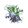
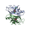
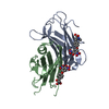
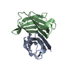
| Structure viewer | Molecule:  Molmil Molmil Jmol/JSmol Jmol/JSmol |
|---|
- Downloads & links
- Download
| PDBx/mmCIF format |  1z7j.cif.gz 1z7j.cif.gz | 63.3 KB | Display |  PDBx/mmCIF format PDBx/mmCIF format |
|---|---|---|---|---|
| PDB format |  pdb1z7j.ent.gz pdb1z7j.ent.gz | 46.9 KB | Display |  PDB format PDB format |
| PDBx/mmJSON format |  1z7j.json.gz 1z7j.json.gz | Tree view |  PDBx/mmJSON format PDBx/mmJSON format | |
| Others |  Other downloads Other downloads |
-Validation report
| Arichive directory |  https://data.pdbj.org/pub/pdb/validation_reports/z7/1z7j https://data.pdbj.org/pub/pdb/validation_reports/z7/1z7j ftp://data.pdbj.org/pub/pdb/validation_reports/z7/1z7j ftp://data.pdbj.org/pub/pdb/validation_reports/z7/1z7j | HTTPS FTP |
|---|
-Related structure data
| Related structure data |  2roxS S: Starting model for refinement |
|---|---|
| Similar structure data |
- Links
Links
- Assembly
| Deposited unit | 
| |||||||||
|---|---|---|---|---|---|---|---|---|---|---|
| 1 | 
| |||||||||
| Unit cell |
| |||||||||
| Components on special symmetry positions |
|
- Components
| #1: Protein | Mass: 13777.360 Da / Num. of mol.: 2 / Source method: isolated from a natural source / Source: (natural)  Homo sapiens (human) / Tissue: PLASMA / References: UniProt: P02766 Homo sapiens (human) / Tissue: PLASMA / References: UniProt: P02766#2: Chemical | #3: Water | ChemComp-HOH / | |
|---|
-Experimental details
-Experiment
| Experiment | Method:  X-RAY DIFFRACTION / Number of used crystals: 1 X-RAY DIFFRACTION / Number of used crystals: 1 |
|---|
- Sample preparation
| Crystal | Density Matthews: 2.26 Å3/Da / Density % sol: 41.7 % |
|---|---|
| Crystal grow | Temperature: 277 K / Method: vapor diffusion, hanging drop / pH: 5.5 Details: 48% ammonium sulfate, 0.l M phosphate buffer, pH 5.50, VAPOR DIFFUSION, HANGING DROP, temperature 4.0K |
-Data collection
| Diffraction | Mean temperature: 293 K |
|---|---|
| Diffraction source | Source:  ROTATING ANODE / Type: RIGAKU RU200 / Wavelength: 1.5418 Å ROTATING ANODE / Type: RIGAKU RU200 / Wavelength: 1.5418 Å |
| Detector | Type: RIGAKU RAXIS II / Detector: IMAGE PLATE / Date: Jan 22, 1999 / Details: COLLIMATOR |
| Radiation | Monochromator: GRAPHITE / Protocol: SINGLE WAVELENGTH / Monochromatic (M) / Laue (L): M / Scattering type: x-ray |
| Radiation wavelength | Wavelength: 1.5418 Å / Relative weight: 1 |
| Reflection | Resolution: 2.2→43.461 Å / Num. obs: 11346 / % possible obs: 94.5 % / Observed criterion σ(F): 0 / Observed criterion σ(I): 1 / Redundancy: 3.45 % / Biso Wilson estimate: 23.3 Å2 / Rmerge(I) obs: 0.0587 / Net I/σ(I): 15.2777 |
| Reflection shell | Resolution: 2.2→2.3 Å / Redundancy: 2.37 % / Rmerge(I) obs: 0.061 / Mean I/σ(I) obs: 2.44 / % possible all: 73.7 |
- Processing
| Software |
| ||||||||||||||||||||||||||||||||||||
|---|---|---|---|---|---|---|---|---|---|---|---|---|---|---|---|---|---|---|---|---|---|---|---|---|---|---|---|---|---|---|---|---|---|---|---|---|---|
| Refinement | Method to determine structure:  FOURIER SYNTHESIS FOURIER SYNTHESISStarting model: DIMER GENERATED FROM PDB ENTRY 2ROX STRUCTURE WITH ONLY PROTEIN ATOMS FROM RESIDUES 10-125 INCLUDED Resolution: 2.2→14.87 Å / Rfactor Rfree error: 0.007 / Isotropic thermal model: RESTRAINED / Cross valid method: THROUGHOUT / σ(F): 2 / Stereochemistry target values: Engh & Huber Details: THIS COORDINATE SET COMPRISES TWO MONOMERS OF HUMAN TTR DIMER (CHAINS A AND B). MULTIPLE CONFORMATIONS HAVE BEEN FOUND FOR 13 RESIDUES. FOUR LIGAND MOLECULES HAVE BEEN FOUND (3,3',5,5'- ...Details: THIS COORDINATE SET COMPRISES TWO MONOMERS OF HUMAN TTR DIMER (CHAINS A AND B). MULTIPLE CONFORMATIONS HAVE BEEN FOUND FOR 13 RESIDUES. FOUR LIGAND MOLECULES HAVE BEEN FOUND (3,3',5,5'-TETRAIODOTHYROACETIC ACID) IN BOTH THE FORWARD AND THE REVERSE ORIENTATION. THERE ARE 81 WATER MOLECULES INCLUDED IN THE MODEL. RESIDUES A1-A9 AND A126-A127 OF THE FIRST MONOMER AND RESIDUES B201-B207 AND B326-B327 FROM THE SECOND ARE ILL-DEFINED IN THE ELECTRON DENSITY MAPS WEIGHTED ML MAPS AND HAVE BEEN OMITTED.
| ||||||||||||||||||||||||||||||||||||
| Solvent computation | Solvent model: FLAT MODEL / Bsol: 42.14 Å2 / ksol: 0.29 e/Å3 | ||||||||||||||||||||||||||||||||||||
| Displacement parameters | Biso mean: 32.5 Å2
| ||||||||||||||||||||||||||||||||||||
| Refine analyze |
| ||||||||||||||||||||||||||||||||||||
| Refinement step | Cycle: LAST / Resolution: 2.2→14.87 Å
| ||||||||||||||||||||||||||||||||||||
| Refine LS restraints |
| ||||||||||||||||||||||||||||||||||||
| LS refinement shell | Resolution: 2.2→2.3 Å / Rfactor Rfree error: 0.033 / Total num. of bins used: 8
| ||||||||||||||||||||||||||||||||||||
| Xplor file |
|
 Movie
Movie Controller






 PDBj
PDBj


