 Movie
Movie Controller


+ Open data

- Basic information
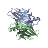
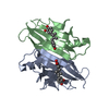
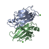
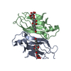
| Entry | Database: PDB / ID: 1f41 | ||||||
|---|---|---|---|---|---|---|---|
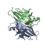
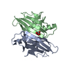
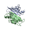
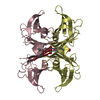
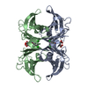
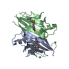
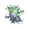
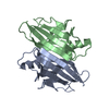
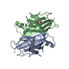
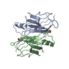
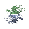
| Title | CRYSTAL STRUCTURE OF HUMAN TRANSTHYRETIN AT 1.5A RESOLUTION | ||||||
 Components Components | TRANSTHYRETIN | ||||||
 Keywords Keywords | TRANSPORT PROTEIN / Greek key beta barrel | ||||||
| Function / homology |  Function and homology information Function and homology informationDefective visual phototransduction due to STRA6 loss of function / negative regulation of glomerular filtration / The canonical retinoid cycle in rods (twilight vision) / purine nucleobase metabolic process / hormone binding / Non-integrin membrane-ECM interactions / phototransduction, visible light / molecular sequestering activity / retinoid metabolic process / Retinoid metabolism and transport ...Defective visual phototransduction due to STRA6 loss of function / negative regulation of glomerular filtration / The canonical retinoid cycle in rods (twilight vision) / purine nucleobase metabolic process / hormone binding / Non-integrin membrane-ECM interactions / phototransduction, visible light / molecular sequestering activity / retinoid metabolic process / Retinoid metabolism and transport / hormone activity / azurophil granule lumen / Amyloid fiber formation / Neutrophil degranulation / protein-containing complex binding / protein-containing complex / : / extracellular exosome / extracellular region / identical protein binding Similarity search - Function | ||||||
| Biological species |  Homo sapiens (human) Homo sapiens (human) | ||||||
| Method |  X-RAY DIFFRACTION / SYNCHROTRON / Resolution: 1.3 Å X-RAY DIFFRACTION / SYNCHROTRON / Resolution: 1.3 Å | ||||||
 Authors Authors | Hornberg, A. / Eneqvist, T. / Olofsson, A. / Lundgren, E. / Sauer-Eriksson, A.E. | ||||||
 Citation Citation | Journal: J.Mol.Biol. / Year: 2000 Title: A comparative analysis of 23 structures of the amyloidogenic protein transthyretin. Authors: Hornberg, A. / Eneqvist, T. / Olofsson, A. / Lundgren, E. / Sauer-Eriksson, A.E. | ||||||
| History |
|
- Structure visualization
| Structure viewer | Molecule: MolmilJmol/JSmol |
|---|
- Downloads & links
-Download
| PDBx/mmCIF format | 1f41.cif.gz | 60.8 KB | Display | PDBx/mmCIF format |
|---|---|---|---|---|
| PDB format | pdb1f41.ent.gz | 46 KB | Display | PDB format |
| PDBx/mmJSON format | 1f41.json.gz | Tree view | PDBx/mmJSON format | |
| Others |  Other downloads Other downloads |
-Validation report
| Arichive directory | https://data.pdbj.org/pub/pdb/validation_reports/f4/1f41ftp://data.pdbj.org/pub/pdb/validation_reports/f4/1f41 | HTTPS FTP |
|---|
-Related structure data
| Similar structure data |
|---|


-Links
 PDBj
PDBj


- Assembly
| Deposited unit | 
| |||||||||
|---|---|---|---|---|---|---|---|---|---|---|
| 1 | 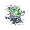
| |||||||||
| Unit cell |
| |||||||||
| Components on special symmetry positions |
| |||||||||
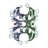
| Details | The biological assembly is a tetramer constructed from the AB dimer and a symmetry dimer generated by the two-fold. |
-Components
| #1: Protein | Mass: 13777.360 Da / Num. of mol.: 2 Source method: isolated from a genetically manipulated source Source: (gene. exp.) Homo sapiens (human) / Tissue fraction: PLASMA / Plasmid: PET3A / Production host:  #2: Water | ChemComp-HOH / |  Mass: 18.015 Da / Num. of mol.: 186 / Source method: isolated from a natural source / Formula: H2O Mass: 18.015 Da / Num. of mol.: 186 / Source method: isolated from a natural source / Formula: H2O |
|---|
-Experimental details
-Experiment
| Experiment | Method: X-RAY DIFFRACTION / Number of used crystals: 1 |
|---|
- Sample preparation
| Crystal | Density Matthews: 2.12 Å3/Da / Density % sol: 67.8 % | |||||||||||||||||||||||||||||||||||
|---|---|---|---|---|---|---|---|---|---|---|---|---|---|---|---|---|---|---|---|---|---|---|---|---|---|---|---|---|---|---|---|---|---|---|---|---|
| Crystal grow | Temperature: 293 K / Method: vapor diffusion, hanging drop / pH: 7 Details: 40% PEG 550MME, 0.1M HEPES, pH 7.0, VAPOR DIFFUSION, HANGING DROP, temperature 293.0K | |||||||||||||||||||||||||||||||||||
| Crystal grow | *PLUS Temperature: 20 ℃ / pH: 7.5 | |||||||||||||||||||||||||||||||||||
| Components of the solutions | *PLUS
|
-Data collection
| Diffraction | Mean temperature: 100 K |
|---|---|
| Diffraction source | Source: SYNCHROTRON / Site: MAX II  / Beamline: I711 / Wavelength: 1 / Beamline: I711 / Wavelength: 1 |
| Detector | Type: MARRESEARCH / Detector: IMAGE PLATE / Date: Dec 13, 1998 |
| Radiation | Protocol: SINGLE WAVELENGTH / Monochromatic (M) / Laue (L): M / Scattering type: x-ray |
| Radiation wavelength | Wavelength: 1 Å / Relative weight: 1 |
| Reflection | Resolution: 1.5→20 Å / Num. all: 439563 / Num. obs: 38636 / % possible obs: 97.1 % / Observed criterion σ(F): 0 / Observed criterion σ(I): 1 / Redundancy: 9.5 % / Biso Wilson estimate: 17.4 Å2 / Rmerge(I) obs: 0.05 / Net I/σ(I): 21 |
| Reflection shell | Resolution: 1.5→1.54 Å / Redundancy: 5.8 % / Rmerge(I) obs: 0.174 / Num. unique all: 2797 / % possible all: 95.7 |
| Reflection | *PLUS Num. obs: 58852 / Num. measured all: 439563 / Rmerge(I) obs: 0.05 |
| Reflection shell | *PLUS % possible obs: 95.7 % / Mean I/σ(I) obs: 9.6 |
- Processing
| Software |
| |||||||||||||||||||||||||
|---|---|---|---|---|---|---|---|---|---|---|---|---|---|---|---|---|---|---|---|---|---|---|---|---|---|---|
| Refinement | Resolution: 1.3→20 Å / σ(F): 0 / σ(I): 0 / Stereochemistry target values: Engh & Huber Details: Individual anisotropic B-factor refinement as implemented in Refmac was performed. Reflections to 1.3A resolution (40% of total) are also included in the refinement. The completeness at 1.5A ...Details: Individual anisotropic B-factor refinement as implemented in Refmac was performed. Reflections to 1.3A resolution (40% of total) are also included in the refinement. The completeness at 1.5A is 97.9%. Crystals were grown at physiological pH. CNS was also used for refinement.
| |||||||||||||||||||||||||
| Refinement step | Cycle: LAST / Resolution: 1.3→20 Å
| |||||||||||||||||||||||||
| Refine LS restraints |
| |||||||||||||||||||||||||
| Software | *PLUS Name: REFMAC / Classification: refinement | |||||||||||||||||||||||||
| Refinement | *PLUS Highest resolution: 1.3 Å / Lowest resolution: 20 Å / σ(F): 0 / % reflection Rfree: 10 % | |||||||||||||||||||||||||
| Solvent computation | *PLUS | |||||||||||||||||||||||||
| Displacement parameters | *PLUS | |||||||||||||||||||||||||
| Refine LS restraints | *PLUS
| |||||||||||||||||||||||||
| LS refinement shell | *PLUS Highest resolution: 1.3 Å / Lowest resolution: 1.36 Å / Rfactor Rfree: 0.271 / Num. reflection Rfree: 290 / Rfactor Rwork: 0.252 / Num. reflection Rwork: 2983 |
