Mass: 18.015 Da / Num. of mol.: 18 / Source method: isolated from a natural source / Formula: H2O
-
Details
Has protein modification
Y
-
Experimental details
-
Experiment
Experiment
Method: X-RAY DIFFRACTION / Number of used crystals: 1
-
Sample preparation
Crystal
Density Matthews: 3.17 Å3/Da / Density % sol: 61.19 % Description: DATA WERE COLLECTED USING THE OSCILLATION METHOD
Crystal grow
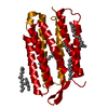
Method: vapor diffusion / pH: 5.2 Details: THE CRYSTALLIZATION CONSISTS OF TWO STEPS. FIRST, A MIXTURE OF 5 MG/ML PURPLE MEMBRANE, 0.25% OTG, 1 M AMMONIUM SULFATE, 0.16 M NACL, 0.04 M SODIUM CITRATE (PH5.2) , 0.04% NAN3 WAS INCUBATED ...Details: THE CRYSTALLIZATION CONSISTS OF TWO STEPS. FIRST, A MIXTURE OF 5 MG/ML PURPLE MEMBRANE, 0.25% OTG, 1 M AMMONIUM SULFATE, 0.16 M NACL, 0.04 M SODIUM CITRATE (PH5.2) , 0.04% NAN3 WAS INCUBATED AND 15% TREHALOSE AT 305K FOR 5 DAYS. THIS RESULTED IN THE FORMATION OF SPHERICAL VESICLES WITH A DIAMETER OF 50 NM. AFTER SEDIMENTAL MATERIALS WERE REMOVED BY CENTRIFUGATION (4000G X 10 MIN), A SUSPENSION OF THE SPHERICAL VESICLES WAS COOLED TO 278K AND CONCENTRATED BY VAPOR DIFFUSION AGAINST A RESERVOIR SOLUTION CONTAINING 2.0 M AMMONIUM SULFATE 0.08M SODIUM CITRATE (PH 5.2) AND 30% TREHALOSE. INCUBATION FOR A COUPLE OF MONTHS YIELDED HEXAGONAL CRYSTALS.
In the structure databanks used in Yorodumi, some data are registered as the other names, "COVID-19 virus" and "2019-nCoV". Here are the details of the virus and the list of structure data.
Jan 31, 2019. EMDB accession codes are about to change! (news from PDBe EMDB page)
EMDB accession codes are about to change! (news from PDBe EMDB page)
The allocation of 4 digits for EMDB accession codes will soon come to an end. Whilst these codes will remain in use, new EMDB accession codes will include an additional digit and will expand incrementally as the available range of codes is exhausted. The current 4-digit format prefixed with “EMD-” (i.e. EMD-XXXX) will advance to a 5-digit format (i.e. EMD-XXXXX), and so on. It is currently estimated that the 4-digit codes will be depleted around Spring 2019, at which point the 5-digit format will come into force.
The EM Navigator/Yorodumi systems omit the EMD- prefix.
Related info.:Q: What is EMD? / ID/Accession-code notation in Yorodumi/EM Navigator
Yorodumi is a browser for structure data from EMDB, PDB, SASBDB, etc.
This page is also the successor to EM Navigator detail page, and also detail information page/front-end page for Omokage search.
The word "yorodu" (or yorozu) is an old Japanese word meaning "ten thousand". "mi" (miru) is to see.
Related info.:EMDB / PDB / SASBDB / Comparison of 3 databanks / Yorodumi Search / Aug 31, 2016. New EM Navigator & Yorodumi / Yorodumi Papers / Jmol/JSmol / Function and homology information / Changes in new EM Navigator and Yorodumi
 Movie
Movie Controller

 Yorodumi
 Open data
 Basic information
 Components
 Keywords
 Function and homology information
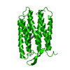
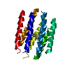
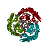
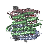
 HALOBACTERIUM SALINARIUM (Halophile)
 X-RAY DIFFRACTION /
  Authors
 Citation
 Structure visualization
 Downloads & links
 Other downloads


 PDBj
PDBj


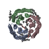
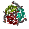
 Assembly


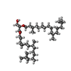

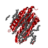
 Mass: 284.436 Da / Num. of mol.: 1 / Source method: obtained synthetically / Formula: C20H28O
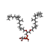
 Mass: 733.137 Da / Num. of mol.: 1 / Source method: obtained synthetically / Formula: C43H89O6P
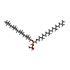
 Mass: 653.157 Da / Num. of mol.: 2 / Source method: obtained synthetically / Formula: C43H88O3
 Mass: 885.179 Da / Num. of mol.: 1 / Source method: obtained synthetically / Formula: C46H94O11P2
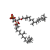
 Mass: 807.215 Da / Num. of mol.: 1 / Source method: obtained synthetically / Formula: C46H95O8P
 Sample preparation
 / Beamline: BL44B2 / Wavelength: 0.7
  Processing