 ムービー
ムービー コントローラー


+ データを開く

- 基本情報
基本情報
| 登録情報 | データベース: PDB / ID: 1d7m | ||||||
|---|---|---|---|---|---|---|---|
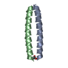
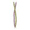
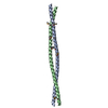
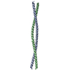
| タイトル | COILED-COIL DIMERIZATION DOMAIN FROM CORTEXILLIN I | ||||||
 要素 要素 | CORTEXILLIN I | ||||||
 キーワード キーワード | CONTRACTILE PROTEIN / COILED-COIL / COILED-COIL TRIGGER SITE / ALPHA HELIX / DIMERIZATION | ||||||
| 機能・相同性 |  機能・相同性情報 機能・相同性情報lateral part of motile cell / sorocarp stalk morphogenesis / negative regulation of protein localization to cell cortex / actomyosin contractile ring contraction / regulation of myosin II filament assembly / protein localization to cleavage furrow / chemotaxis to cAMP / cell trailing edge / aggregation involved in sorocarp development / positive regulation of cytoskeleton organization ...lateral part of motile cell / sorocarp stalk morphogenesis / negative regulation of protein localization to cell cortex / actomyosin contractile ring contraction / regulation of myosin II filament assembly / protein localization to cleavage furrow / chemotaxis to cAMP / cell trailing edge / aggregation involved in sorocarp development / positive regulation of cytoskeleton organization / adenylate cyclase-activating G protein-coupled cAMP receptor signaling pathway / actomyosin contractile ring / mitotic spindle midzone / basal cortex / actin crosslink formation / alpha-catenin binding / cleavage furrow formation / detection of mechanical stimulus / bleb assembly / protein kinase regulator activity / cortical actin cytoskeleton organization / cortical actin cytoskeleton / cell leading edge / cleavage furrow / mitotic cytokinesis / actin filament bundle assembly / response to mechanical stimulus / phagocytic vesicle / cell projection / phosphatidylinositol-4,5-bisphosphate binding / response to bacterium / cell morphogenesis / actin filament binding / intracellular protein localization / actin cytoskeleton / regulation of gene expression / actin cytoskeleton organization / cell cortex / vesicle / positive regulation of gene expression / identical protein binding / plasma membrane / cytosol 類似検索 - 分子機能 | ||||||
| 生物種 |  | ||||||
| 手法 |  X線回折 / シンクロトロン / 解像度: 2.7 Å X線回折 / シンクロトロン / 解像度: 2.7 Å | ||||||
 データ登録者 データ登録者 | Burkhard, P. / Kammerer, R.A. / Steinmetz, M.O. / Bourenkov, G.P. / Aebi, U. | ||||||
 引用 引用 | ジャーナル: Structure Fold.Des. / 年: 2000 タイトル: The coiled-coil trigger site of the rod domain of cortexillin I unveils a distinct network of interhelical and intrahelical salt bridges. 著者: Burkhard, P. / Kammerer, R.A. / Steinmetz, M.O. / Bourenkov, G.P. / Aebi, U. #1: ジャーナル: J.Struct.Biol. / 年: 1998タイトル: Crystallization and Preliminary X-Ray Diffraction Analysis of the 190 A Long Coiled-Coil Dimerization Domain of the Actin-Bundling Protein Cortexillin I from Dictyostelium discoideum 著者: Burkhard, P. / Steinmetz, M.O. / Schulthess, T. / Landwehr, R. / Aebi, U. / Kammerer, R.A. | ||||||
| 履歴 |
|
- 構造の表示
構造の表示
| 構造ビューア | 分子: MolmilJmol/JSmol |
|---|
- ダウンロードとリンク
-ダウンロード
| PDBx/mmCIF形式 | 1d7m.cif.gz | 51.7 KB | 表示 | PDBx/mmCIF形式 |
|---|---|---|---|---|
| PDB形式 | pdb1d7m.ent.gz | 39.5 KB | 表示 | PDB形式 |
| PDBx/mmJSON形式 | 1d7m.json.gz | ツリー表示 | PDBx/mmJSON形式 | |
| その他 |  その他のダウンロード その他のダウンロード |
-検証レポート
| アーカイブディレクトリ | https://data.pdbj.org/pub/pdb/validation_reports/d7/1d7mftp://data.pdbj.org/pub/pdb/validation_reports/d7/1d7m | HTTPS FTP |
|---|
-関連構造データ
| 関連構造データ | |
|---|---|
| 類似構造データ |

-リンク
 PDBj
PDBj- 集合体
集合体
| 登録構造単位 | 
| ||||||||
|---|---|---|---|---|---|---|---|---|---|
| 1 |
| ||||||||
| 2 | 
| ||||||||
| 単位格子 |
| ||||||||
| Components on special symmetry positions |
|
-要素
| #1: タンパク質 | 分子量: 11623.278 Da / 分子数: 2 / 断片: COILED-COIL DIMERIZATION DOMAIN / 由来タイプ: 組換発現 由来: (組換発現) 発現宿主:  #2: 水 | ChemComp-HOH / |  分子量: 18.015 Da / 分子数: 93 / 由来タイプ: 天然 / 式: H2O 分子量: 18.015 Da / 分子数: 93 / 由来タイプ: 天然 / 式: H2O |
|---|
-実験情報
-実験
| 実験 | 手法: X線回折 / 使用した結晶の数: 2 |
|---|
- 試料調製
試料調製
| 結晶 | マシュー密度: 4.46 Å3/Da / 溶媒含有率: 72.43 % | ||||||||||||||||||||||||||||||||||||||||||||||||
|---|---|---|---|---|---|---|---|---|---|---|---|---|---|---|---|---|---|---|---|---|---|---|---|---|---|---|---|---|---|---|---|---|---|---|---|---|---|---|---|---|---|---|---|---|---|---|---|---|---|
| 結晶化 | 温度: 297 K / 手法: 蒸気拡散法, ハンギングドロップ法 / pH: 6.5 詳細: 1.5 M AMMONIUM SULFATE 100 MM TRIS (PH 6.5) 2.5 % PEG 400 1.0 % DIOXANE, VAPOR DIFFUSION, HANGING DROP, temperature 297K | ||||||||||||||||||||||||||||||||||||||||||||||||
| 結晶化 | *PLUS pH: 7.4 / 詳細: Burkhard, P., (1998) J.Struct.Biol., 122, 293. | ||||||||||||||||||||||||||||||||||||||||||||||||
| 溶液の組成 | *PLUS
|
-データ収集
| 回折 |
| |||||||||||||||
|---|---|---|---|---|---|---|---|---|---|---|---|---|---|---|---|---|
| 放射光源 |
| |||||||||||||||
| 検出器 |
| |||||||||||||||
| 放射 |
| |||||||||||||||
| 放射波長 |
| |||||||||||||||
| 反射 | 解像度: 2.7→30 Å / Num. all: 31909 / Num. obs: 11302 / % possible obs: 95.9 % / 冗長度: 2.8 % / Biso Wilson estimate: 37.9 Å2 / Rmerge(I) obs: 0.111 / Net I/σ(I): 5.3 | |||||||||||||||
| 反射 シェル | 解像度: 2.7→2.85 Å / 冗長度: 2.7 % / Rmerge(I) obs: 0.462 / % possible all: 88.2 | |||||||||||||||
| 反射 | *PLUS % possible obs: 94.6 % / Num. measured all: 49645 / Rmerge(I) obs: 0.078 |

- 解析
解析
| ソフトウェア |
| ||||||||||||||||||||||||||||||||||||||||||||||||||||||||||||||||||||||||||||||||
|---|---|---|---|---|---|---|---|---|---|---|---|---|---|---|---|---|---|---|---|---|---|---|---|---|---|---|---|---|---|---|---|---|---|---|---|---|---|---|---|---|---|---|---|---|---|---|---|---|---|---|---|---|---|---|---|---|---|---|---|---|---|---|---|---|---|---|---|---|---|---|---|---|---|---|---|---|---|---|---|---|---|
| 精密化 | 解像度: 2.7→37.3 Å / Rfactor Rfree error: 0.011 / Data cutoff high absF: 1675937.19 / Data cutoff high rms absF: 1675937.19 / Data cutoff low absF: 0 / Isotropic thermal model: CONSTRAINED / 交差検証法: THROUGHOUT / σ(F): 3
| ||||||||||||||||||||||||||||||||||||||||||||||||||||||||||||||||||||||||||||||||
| 溶媒の処理 | 溶媒モデル: FLAT MODEL / Bsol: 71.69 Å2 / ksol: 0.406 e/Å3 | ||||||||||||||||||||||||||||||||||||||||||||||||||||||||||||||||||||||||||||||||
| 原子変位パラメータ | Biso mean: 56.9 Å2
| ||||||||||||||||||||||||||||||||||||||||||||||||||||||||||||||||||||||||||||||||
| Refine analyze |
| ||||||||||||||||||||||||||||||||||||||||||||||||||||||||||||||||||||||||||||||||
| 精密化ステップ | サイクル: LAST / 解像度: 2.7→37.3 Å
| ||||||||||||||||||||||||||||||||||||||||||||||||||||||||||||||||||||||||||||||||
| 拘束条件 |
| ||||||||||||||||||||||||||||||||||||||||||||||||||||||||||||||||||||||||||||||||
| Refine LS restraints NCS | NCS model details: CONSTRAINED | ||||||||||||||||||||||||||||||||||||||||||||||||||||||||||||||||||||||||||||||||
| LS精密化 シェル | 解像度: 2.7→2.87 Å / Rfactor Rfree error: 0.052 / Total num. of bins used: 6
| ||||||||||||||||||||||||||||||||||||||||||||||||||||||||||||||||||||||||||||||||
| Xplor file |
| ||||||||||||||||||||||||||||||||||||||||||||||||||||||||||||||||||||||||||||||||
| ソフトウェア | *PLUS 名称: CNS / バージョン: 0.9 / 分類: refinement | ||||||||||||||||||||||||||||||||||||||||||||||||||||||||||||||||||||||||||||||||
| 精密化 | *PLUS 最低解像度: 30 Å / σ(F): 3 / % reflection Rfree: 5 % / Rfactor obs: 0.218 / Rfactor Rfree: 0.257 | ||||||||||||||||||||||||||||||||||||||||||||||||||||||||||||||||||||||||||||||||
| 溶媒の処理 | *PLUS | ||||||||||||||||||||||||||||||||||||||||||||||||||||||||||||||||||||||||||||||||
| 原子変位パラメータ | *PLUS Biso mean: 56.9 Å2 | ||||||||||||||||||||||||||||||||||||||||||||||||||||||||||||||||||||||||||||||||
| 拘束条件 | *PLUS
| ||||||||||||||||||||||||||||||||||||||||||||||||||||||||||||||||||||||||||||||||
| LS精密化 シェル | *PLUS Rfactor Rfree: 0.381 / % reflection Rfree: 5.1 % / Rfactor Rwork: 0.268 |
