 Movie
Movie Controller

[English] 日本語
 Yorodumi
- PDB-3u1c: Anti-parallel dimer of N-terminal 98-aa fragment of smooth muscle... -
+ Open data

- Basic information
| Entry | Database: PDB / ID: 3u1c | ||||||
|---|---|---|---|---|---|---|---|
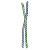
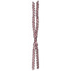
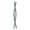
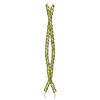
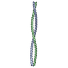
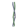
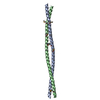
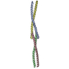
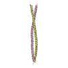
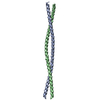
| Title | Anti-parallel dimer of N-terminal 98-aa fragment of smooth muscle tropomyosin alpha | ||||||
 Components Components | Tropomyosin alpha-1 chain | ||||||
 Keywords Keywords | CONTRACTILE PROTEIN / Anti-parallel coiled coil | ||||||
| Function / homology |  Function and homology information Function and homology informationStriated Muscle Contraction / Smooth Muscle Contraction / cardiac muscle contraction / cytoskeletal protein binding / actin filament organization / actin filament / actin filament binding / actin cytoskeleton / protein heterodimerization activity / protein homodimerization activity ...Striated Muscle Contraction / Smooth Muscle Contraction / cardiac muscle contraction / cytoskeletal protein binding / actin filament organization / actin filament / actin filament binding / actin cytoskeleton / protein heterodimerization activity / protein homodimerization activity / identical protein binding / plasma membrane Similarity search - Function | ||||||
| Biological species |  | ||||||
| Method |  X-RAY DIFFRACTION / SYNCHROTRON / SAD / Resolution: 1.8 Å X-RAY DIFFRACTION / SYNCHROTRON / SAD / Resolution: 1.8 Å | ||||||
 Authors Authors | Jampani, N. / Dominguez, R. | ||||||
 Citation Citation | Journal: J.Biol.Chem. / Year: 2012 Title: Structural analysis of smooth muscle tropomyosin alpha and beta isoforms. Authors: Rao, J.N. / Rivera-Santiago, R. / Li, X.E. / Lehman, W. / Dominguez, R. | ||||||
| History |
|
- Structure visualization
| Structure viewer | Molecule: MolmilJmol/JSmol |
|---|
- Downloads & links
-Download
| PDBx/mmCIF format | 3u1c.cif.gz | 96.7 KB | Display | PDBx/mmCIF format |
|---|---|---|---|---|
| PDB format | pdb3u1c.ent.gz | 76.5 KB | Display | PDB format |
| PDBx/mmJSON format | 3u1c.json.gz | Tree view | PDBx/mmJSON format | |
| Others |  Other downloads Other downloads |
-Validation report
| Arichive directory | https://data.pdbj.org/pub/pdb/validation_reports/u1/3u1cftp://data.pdbj.org/pub/pdb/validation_reports/u1/3u1c | HTTPS FTP |
|---|
-Related structure data


-Links
 PDBj
PDBj- Assembly
| Deposited unit | 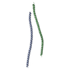
| |||||||||||||||
|---|---|---|---|---|---|---|---|---|---|---|---|---|---|---|---|---|
| 1 | 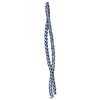
| |||||||||||||||
| 2 | 
| |||||||||||||||
| Unit cell |
| |||||||||||||||
| Components on special symmetry positions |
|
-Components
| #1: Protein | Mass: 11595.910 Da / Num. of mol.: 2 / Fragment: unp residues 1-98 Source method: isolated from a genetically manipulated source Source: (gene. exp.)  #2: Water | ChemComp-HOH / |  Mass: 18.015 Da / Num. of mol.: 149 / Source method: isolated from a natural source / Formula: H2O Mass: 18.015 Da / Num. of mol.: 149 / Source method: isolated from a natural source / Formula: H2O |
|---|
-Experimental details
-Experiment
| Experiment | Method: X-RAY DIFFRACTION / Number of used crystals: 2 |
|---|
- Sample preparation
| Crystal | Density Matthews: 2.52 Å3/Da / Density % sol: 51.26 % |
|---|---|
| Crystal grow | Temperature: 293 K / Method: vapor diffusion, hanging drop / pH: 4 Details: 0.1 M Sodium acetate, pH 4.0, 40 mM Calcium chloride, 32-35 % 2-METHYL-2,4-PENTANEDIOL, VAPOR DIFFUSION, HANGING DROP, temperature 293K |
-Data collection
| Diffraction |
| ||||||||||||||||||
|---|---|---|---|---|---|---|---|---|---|---|---|---|---|---|---|---|---|---|---|
| Diffraction source |
| ||||||||||||||||||
| Detector |
| ||||||||||||||||||
| Radiation |
| ||||||||||||||||||
| Radiation wavelength |
| ||||||||||||||||||
| Reflection | Resolution: 1.75→25 Å / Num. all: 24608 / Num. obs: 23590 / % possible obs: 95.9 % / Redundancy: 17.76 % / Rmerge(I) obs: 0.0757 / Net I/σ(I): 24.9 | ||||||||||||||||||
| Reflection shell | Resolution: 1.75→1.85 Å / Redundancy: 12.69 % / Rmerge(I) obs: 0.4364 / Mean I/σ(I) obs: 4.59 / Num. unique all: 3675 / % possible all: 94.8 |

- Processing
| Software |
| |||||||||||||||||||||||||||||||||||||||||||||||||||||||||||||||||||||||||||
|---|---|---|---|---|---|---|---|---|---|---|---|---|---|---|---|---|---|---|---|---|---|---|---|---|---|---|---|---|---|---|---|---|---|---|---|---|---|---|---|---|---|---|---|---|---|---|---|---|---|---|---|---|---|---|---|---|---|---|---|---|---|---|---|---|---|---|---|---|---|---|---|---|---|---|---|---|
| Refinement | Method to determine structure: SAD / Resolution: 1.8→25 Å / SU ML: 0.26 / σ(F): 0 / Phase error: 31.53 / Stereochemistry target values: ML
| |||||||||||||||||||||||||||||||||||||||||||||||||||||||||||||||||||||||||||
| Solvent computation | Shrinkage radii: 0.72 Å / VDW probe radii: 1 Å / Solvent model: FLAT BULK SOLVENT MODEL / Bsol: 54.254 Å2 / ksol: 0.378 e/Å3 | |||||||||||||||||||||||||||||||||||||||||||||||||||||||||||||||||||||||||||
| Displacement parameters |
| |||||||||||||||||||||||||||||||||||||||||||||||||||||||||||||||||||||||||||
| Refinement step | Cycle: LAST / Resolution: 1.8→25 Å
| |||||||||||||||||||||||||||||||||||||||||||||||||||||||||||||||||||||||||||
| Refine LS restraints |
| |||||||||||||||||||||||||||||||||||||||||||||||||||||||||||||||||||||||||||
| LS refinement shell | Refine-ID: X-RAY DIFFRACTION
| |||||||||||||||||||||||||||||||||||||||||||||||||||||||||||||||||||||||||||
| Refinement TLS params. | Method: refined / Refine-ID: X-RAY DIFFRACTION
| |||||||||||||||||||||||||||||||||||||||||||||||||||||||||||||||||||||||||||
| Refinement TLS group |
|
