- PDB-3tnu: Heterocomplex of coil 2B domains of human intermediate filament p... -
+
Open data
ID or keywords:
Loading...
-
Basic information
Entry
Database: PDB / ID: 3tnu
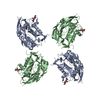
Title
Heterocomplex of coil 2B domains of human intermediate filament proteins, keratin 5 (KRT5) and keratin 14 (KRT14)
Components
Keratin, type I cytoskeletal 14
Keratin, type II cytoskeletal 5
Keywords
CYTOSOLIC PROTEIN / Coiled-coil / Structural Support
Function / homology
Function and homology information
intermediate filament polymerization / intermediate filament bundle assembly / Developmental Lineage of Mammary Gland Luminal Epithelial Cells / hair cycle / structural constituent of skin epidermis / keratin filament / Type I hemidesmosome assembly / keratin filament binding / Keratinization / intermediate filament organization ...intermediate filament polymerization / intermediate filament bundle assembly / Developmental Lineage of Mammary Gland Luminal Epithelial Cells / hair cycle / structural constituent of skin epidermis / keratin filament / Type I hemidesmosome assembly / keratin filament binding / Keratinization / intermediate filament organization / Formation of the cornified envelope / cornified envelope / Developmental Lineage of Mammary Stem Cells / basal part of cell / intermediate filament cytoskeleton / Differentiation of Keratinocytes in Interfollicular Epidermis in Mammalian Skin / intermediate filament / keratinization / Developmental Lineage of Mammary Gland Myoepithelial Cells / epidermis development / response to mechanical stimulus / keratinocyte differentiation / regulation of cell migration / morphogenesis of an epithelium / structural constituent of cytoskeleton / regulation of protein localization / scaffold protein binding / cytoskeleton / extracellular exosome / membrane / nucleus / cytoplasm / cytosol Similarity search - Function
Keratin, type II / Keratin type II head / Keratin type II head / Keratin, type I / Intermediate filament protein, conserved site / Intermediate filament protein / Intermediate filament (IF) rod domain signature. / Intermediate filament, rod domain / Intermediate filament (IF) rod domain profile. / Intermediate filament protein ...Keratin, type II / Keratin type II head / Keratin type II head / Keratin, type I / Intermediate filament protein, conserved site / Intermediate filament protein / Intermediate filament (IF) rod domain signature. / Intermediate filament, rod domain / Intermediate filament (IF) rod domain profile. / Intermediate filament protein / Single alpha-helices involved in coiled-coils or other helix-helix interfaces - #170 / Single alpha-helices involved in coiled-coils or other helix-helix interfaces / Up-down Bundle / Mainly Alpha Similarity search - Domain/homology
Mass: 18.015 Da / Num. of mol.: 6 / Source method: isolated from a natural source / Formula: H2O
Has protein modification
Y
-
Experimental details
-
Experiment
Experiment
Method: X-RAY DIFFRACTION / Number of used crystals: 1
-
Sample preparation
Crystal
Density Matthews: 5.11 Å3/Da / Density % sol: 75.93 %
Crystal grow
Temperature: 293.2 K / Method: vapor diffusion, hanging drop / pH: 8.5 Details: K5/K14 2B protein solution (9 mg/mL) was mixed with an equal volume of reservoir solution containing 2.8M NaCl and 0.1M Tris-HCl (pH 8.5). Small crystals formed and were used to seed the ...Details: K5/K14 2B protein solution (9 mg/mL) was mixed with an equal volume of reservoir solution containing 2.8M NaCl and 0.1M Tris-HCl (pH 8.5). Small crystals formed and were used to seed the same mixture of protein solution/reservoir solution, which yielded larger crystals, VAPOR DIFFUSION, HANGING DROP, temperature 293.2K
In the structure databanks used in Yorodumi, some data are registered as the other names, "COVID-19 virus" and "2019-nCoV". Here are the details of the virus and the list of structure data.
Jan 31, 2019. EMDB accession codes are about to change! (news from PDBe EMDB page)
EMDB accession codes are about to change! (news from PDBe EMDB page)
The allocation of 4 digits for EMDB accession codes will soon come to an end. Whilst these codes will remain in use, new EMDB accession codes will include an additional digit and will expand incrementally as the available range of codes is exhausted. The current 4-digit format prefixed with “EMD-” (i.e. EMD-XXXX) will advance to a 5-digit format (i.e. EMD-XXXXX), and so on. It is currently estimated that the 4-digit codes will be depleted around Spring 2019, at which point the 5-digit format will come into force.
The EM Navigator/Yorodumi systems omit the EMD- prefix.
Related info.:Q: What is EMD? / ID/Accession-code notation in Yorodumi/EM Navigator
Yorodumi is a browser for structure data from EMDB, PDB, SASBDB, etc.
This page is also the successor to EM Navigator detail page, and also detail information page/front-end page for Omokage search.
The word "yorodu" (or yorozu) is an old Japanese word meaning "ten thousand". "mi" (miru) is to see.
Related info.:EMDB / PDB / SASBDB / Comparison of 3 databanks / Yorodumi Search / Aug 31, 2016. New EM Navigator & Yorodumi / Yorodumi Papers / Jmol/JSmol / Function and homology information / Changes in new EM Navigator and Yorodumi
 Movie
Movie Controller

 Yorodumi
 Open data
 Basic information
 Components
 Keywords
 Function and homology information
 Homo sapiens (human)
 X-RAY DIFFRACTION /
  Authors
 Citation
 Structure visualization
 Downloads & links
 Other downloads

 PDBj
PDBj Assembly


 Mass: 18.015 Da / Num. of mol.: 6 / Source method: isolated from a natural source / Formula: H2O
 Sample preparation
 / Beamline: 23-ID-B / Wavelength: 0.97944 Å
 Processing