 Movie
Movie Controller

[English] 日本語
 Yorodumi
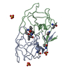
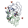
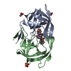
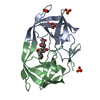
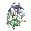
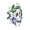
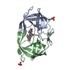
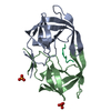
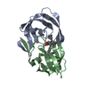
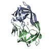
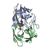
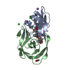
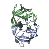
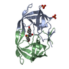
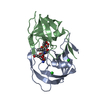
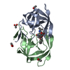
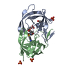
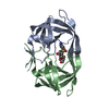
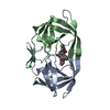
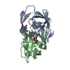
- PDB-1d4k: HIV-1 PROTEASE COMPLEXED WITH A MACROCYCLIC PEPTIDOMIMETIC INHIBITOR -
+ Open data

- Basic information
| Entry | Database: PDB / ID: 1d4k | ||||||
|---|---|---|---|---|---|---|---|
| Title | HIV-1 PROTEASE COMPLEXED WITH A MACROCYCLIC PEPTIDOMIMETIC INHIBITOR | ||||||
 Components Components | HIV-1 PROTEASE | ||||||
 Keywords Keywords | HYDROLASE / HIV / PROTEASE / INHIBITOR / ANTIVIRAL | ||||||
| Function / homology |  Function and homology information Function and homology informationHIV-1 retropepsin / symbiont-mediated activation of host apoptosis / retroviral ribonuclease H / exoribonuclease H / exoribonuclease H activity / DNA integration / viral genome integration into host DNA / establishment of integrated proviral latency / RNA-directed DNA polymerase / RNA stem-loop binding ...HIV-1 retropepsin / symbiont-mediated activation of host apoptosis / retroviral ribonuclease H / exoribonuclease H / exoribonuclease H activity / DNA integration / viral genome integration into host DNA / establishment of integrated proviral latency / RNA-directed DNA polymerase / RNA stem-loop binding / viral penetration into host nucleus / host multivesicular body / RNA-directed DNA polymerase activity / RNA-DNA hybrid ribonuclease activity / Transferases; Transferring phosphorus-containing groups; Nucleotidyltransferases / host cell / viral nucleocapsid / DNA recombination / DNA-directed DNA polymerase / aspartic-type endopeptidase activity / Hydrolases; Acting on ester bonds / DNA-directed DNA polymerase activity / symbiont-mediated suppression of host gene expression / viral translational frameshifting / symbiont entry into host cell / lipid binding / host cell nucleus / host cell plasma membrane / virion membrane / structural molecule activity / proteolysis / DNA binding / zinc ion binding Similarity search - Function | ||||||
| Method |  X-RAY DIFFRACTION / Resolution: 1.85 Å X-RAY DIFFRACTION / Resolution: 1.85 Å | ||||||
 Authors Authors | Tyndall, J.D. / Reid, R.C. / Tyssen, D.P. / Jardine, D.K. / Todd, B. / Passmore, M. / March, D.R. / Pattenden, L.K. / Alewood, D. / Hu, S.H. ...Tyndall, J.D. / Reid, R.C. / Tyssen, D.P. / Jardine, D.K. / Todd, B. / Passmore, M. / March, D.R. / Pattenden, L.K. / Alewood, D. / Hu, S.H. / Alewood, P.F. / Birch, C.J. / Martin, J.L. / Fairlie, D.P. | ||||||
 Citation Citation | Journal: J.Med.Chem. / Year: 2000 Title: Synthesis, stability, antiviral activity, and protease-bound structures of substrate-mimicking constrained macrocyclic inhibitors of HIV-1 protease. Authors: Tyndall, J.D. / Reid, R.C. / Tyssen, D.P. / Jardine, D.K. / Todd, B. / Passmore, M. / March, D.R. / Pattenden, L.K. / Bergman, D.A. / Alewood, D. / Hu, S.H. / Alewood, P.F. / Birch, C.J. / ...Authors: Tyndall, J.D. / Reid, R.C. / Tyssen, D.P. / Jardine, D.K. / Todd, B. / Passmore, M. / March, D.R. / Pattenden, L.K. / Bergman, D.A. / Alewood, D. / Hu, S.H. / Alewood, P.F. / Birch, C.J. / Martin, J.L. / Fairlie, D.P. #1: Journal: Biochemistry / Year: 1999Title: Molecular Recognition of Macrocyclic Peptidomimetic Inhibitors by HIV-1 Protease. Authors: Martin, J.L. / Begun, J. / Schindeler, A. / Wickramasinghe, W.A. / Alewood, D. / Alewood, P.F. / Bergman, D.A. / Brinkworth, R.I. / Abbenante, G. / March, D.R. / Reid, R.C. / Fairlie, D.P. #2: Journal: J.Am.Chem.Soc. / Year: 1996Title: Substrate-Based Cyclic Peptidomimetics Of Phe Ile Val That Inhibit HIV-1 Protease Using a Novel Enzyme Binding Mode Authors: March, D.R. / Abbenante, G. / Bergman, D. / Brinkworth, R.I. / Wickramasinghe, W. / Begun, J. / Martin, J.L. / Fairlie, D.P. #3: Journal: J.Am.Chem.Soc. / Year: 1995Title: Regioselective Structural and Functional Mimicry Of Peptides: Design Of Hydrolytically Stable Cyclic Peptidomimetic Inhibitors Of HIV-1 Protease. Authors: Abbenante, G. / March, D. / Bergman, D. / Hunt, P.A. / Garnham, B. / Dancer, R.J. / Martin, J.L. / Fairlie, D.P. | ||||||
| History |
|
- Structure visualization
| Structure viewer | Molecule: MolmilJmol/JSmol |
|---|
- Downloads & links
-Download
| PDBx/mmCIF format | 1d4k.cif.gz | 51 KB | Display | PDBx/mmCIF format |
|---|---|---|---|---|
| PDB format | pdb1d4k.ent.gz | 40.1 KB | Display | PDB format |
| PDBx/mmJSON format | 1d4k.json.gz | Tree view | PDBx/mmJSON format | |
| Others |  Other downloads Other downloads |
-Validation report
| Arichive directory | https://data.pdbj.org/pub/pdb/validation_reports/d4/1d4kftp://data.pdbj.org/pub/pdb/validation_reports/d4/1d4k | HTTPS FTP |
|---|
-Related structure data


-Links
 PDBj
PDBj

- Assembly
| Deposited unit | 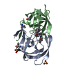
| ||||||||
|---|---|---|---|---|---|---|---|---|---|
| 1 |
| ||||||||
| Unit cell |
|
-Components
| #1: Protein | Mass: 10765.687 Da / Num. of mol.: 2 / Mutation: Q7K, L33I, C67(ABA), C95(ABA) / Source method: obtained synthetically Details: SF2 isolate, chemically synthesised protein corresponds to the protease from HIV-1, with 4 mutations per monomer References: UniProt: P03369, HIV-1 retropepsin #2: Chemical |   Mass: 96.063 Da / Num. of mol.: 2 / Source method: obtained synthetically / Formula: SO4 Mass: 96.063 Da / Num. of mol.: 2 / Source method: obtained synthetically / Formula: SO4#3: Chemical | ChemComp-PI8 / | 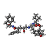  Mass: 698.891 Da / Num. of mol.: 1 / Source method: obtained synthetically / Formula: C41H54N4O6 Mass: 698.891 Da / Num. of mol.: 1 / Source method: obtained synthetically / Formula: C41H54N4O6#4: Water | ChemComp-HOH / |  Mass: 18.015 Da / Num. of mol.: 89 / Source method: isolated from a natural source / Formula: H2O Mass: 18.015 Da / Num. of mol.: 89 / Source method: isolated from a natural source / Formula: H2O |
|---|
-Experimental details
-Experiment
| Experiment | Method: X-RAY DIFFRACTION / Number of used crystals: 1 |
|---|
- Sample preparation
| Crystal | Density Matthews: 2.19 Å3/Da / Density % sol: 43.88 % | ||||||||||||||||||||||||||||||
|---|---|---|---|---|---|---|---|---|---|---|---|---|---|---|---|---|---|---|---|---|---|---|---|---|---|---|---|---|---|---|---|
| Crystal grow | Temperature: 293 K / Method: vapor diffusion, hanging drop / pH: 5.5 Details: ammonium sulfate. acetate buffer, , pH 5.5, VAPOR DIFFUSION, HANGING DROP, temperature 293.0K | ||||||||||||||||||||||||||||||
| Crystal grow | *PLUS Temperature: 20 ℃ / Details: inhibitor:protein molar ratio is 10:1 | ||||||||||||||||||||||||||||||
| Components of the solutions | *PLUS
|
-Data collection
| Diffraction | Mean temperature: 298 K |
|---|---|
| Diffraction source | Source: ROTATING ANODE / Type: RIGAKU RU200 / Wavelength: 1.54 |
| Detector | Type: RIGAKU RAXIS IIC / Detector: IMAGE PLATE / Date: Aug 17, 1998 |
| Radiation | Protocol: SINGLE WAVELENGTH / Monochromatic (M) / Laue (L): M / Scattering type: x-ray |
| Radiation wavelength | Wavelength: 1.54 Å / Relative weight: 1 |
| Reflection | Resolution: 1.85→50 Å / Num. all: 55326 / Num. obs: 51287 / % possible obs: 92.7 % / Observed criterion σ(F): 0 / Observed criterion σ(I): 1 / Redundancy: 3.56 % / Biso Wilson estimate: 13 Å2 / Rmerge(I) obs: 0.051 / Net I/σ(I): 12 |
| Reflection shell | Resolution: 1.85→1.92 Å / Redundancy: 8.19 % / Rmerge(I) obs: 0.277 / Num. unique all: 1457 / % possible all: 87.9 |
| Reflection | *PLUS Num. obs: 15546 / Num. measured all: 51287 |
| Reflection shell | *PLUS % possible obs: 87.9 % |
- Processing
| Software |
| ||||||||||||||||||||||||||||||||||||||||||||||||||||||||||||||||||||||||||||||||
|---|---|---|---|---|---|---|---|---|---|---|---|---|---|---|---|---|---|---|---|---|---|---|---|---|---|---|---|---|---|---|---|---|---|---|---|---|---|---|---|---|---|---|---|---|---|---|---|---|---|---|---|---|---|---|---|---|---|---|---|---|---|---|---|---|---|---|---|---|---|---|---|---|---|---|---|---|---|---|---|---|---|
| Refinement | Resolution: 1.85→8 Å / Rfactor Rfree error: 0.007 / Data cutoff high absF: 10000000 / Data cutoff low absF: 0.001 / Isotropic thermal model: RESTRAINED / Cross valid method: THROUGHOUT / σ(F): 0 / σ(I): 0 / Stereochemistry target values: Engh & Huber
| ||||||||||||||||||||||||||||||||||||||||||||||||||||||||||||||||||||||||||||||||
| Displacement parameters | Biso mean: 24.5 Å2
| ||||||||||||||||||||||||||||||||||||||||||||||||||||||||||||||||||||||||||||||||
| Refine analyze |
| ||||||||||||||||||||||||||||||||||||||||||||||||||||||||||||||||||||||||||||||||
| Refinement step | Cycle: LAST / Resolution: 1.85→8 Å
| ||||||||||||||||||||||||||||||||||||||||||||||||||||||||||||||||||||||||||||||||
| Refine LS restraints |
| ||||||||||||||||||||||||||||||||||||||||||||||||||||||||||||||||||||||||||||||||
| LS refinement shell | Resolution: 1.85→1.96 Å / Rfactor Rfree error: 0.02 / Total num. of bins used: 6
| ||||||||||||||||||||||||||||||||||||||||||||||||||||||||||||||||||||||||||||||||
| Software | *PLUS Name: X-PLOR / Version: 3.851 / Classification: refinement | ||||||||||||||||||||||||||||||||||||||||||||||||||||||||||||||||||||||||||||||||
| Refine LS restraints | *PLUS
| ||||||||||||||||||||||||||||||||||||||||||||||||||||||||||||||||||||||||||||||||
| LS refinement shell | *PLUS Rfactor Rfree: 0.312 / Rfactor Rwork: 0.288 |
