 Movie
Movie Controller

[English] 日本語
 Yorodumi
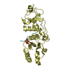
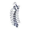
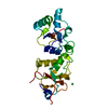
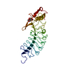
- PDB-1byf: STRUCTURE OF TC14; A C-TYPE LECTIN FROM THE TUNICATE POLYANDROCAR... -
+ Open data

- Basic information
| Entry | Database: PDB / ID: 1byf | ||||||
|---|---|---|---|---|---|---|---|
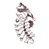
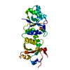
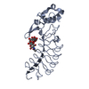
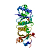
| Title | STRUCTURE OF TC14; A C-TYPE LECTIN FROM THE TUNICATE POLYANDROCARPA MISAKIENSIS | ||||||
 Components Components | PROTEIN (POLYANDROCARPA LECTIN) | ||||||
 Keywords Keywords | SUGAR BINDING PROTEIN / C-TYPE LECTIN / GALACTOSE-SPECIFIC | ||||||
| Function / homology |  Function and homology information Function and homology information | ||||||
| Biological species |  Polyandrocarpa misakiensis (invertebrata) Polyandrocarpa misakiensis (invertebrata) | ||||||
| Method |  X-RAY DIFFRACTION / SIRAS / Resolution: 2 Å X-RAY DIFFRACTION / SIRAS / Resolution: 2 Å | ||||||
 Authors Authors | Poget, S.F. / Legge, G.B. / Bycroft, M. / Williams, R.L. | ||||||
 Citation Citation | Journal: J.Mol.Biol. / Year: 1999 Title: The structure of a tunicate C-type lectin from Polyandrocarpa misakiensis complexed with D -galactose. Authors: Poget, S.F. / Legge, G.B. / Proctor, M.R. / Butler, P.J. / Bycroft, M. / Williams, R.L. #1: Journal: Roux's Arch.Dev.Biol. / Year: 1995Title: Expression of Genes for 2 C-Type Lectins During Budding of the Ascidian Polyandrocarpa-Misakiensis Authors: Shimada, M. / Fujiwara, S. / Kawamura, K. #2: Journal: DEVELOPMENT / Year: 1991Title: Budding-Specific Lectin Induced in Epithelial Cells is an Extracellular Matrix Component for Stem Cell Aggregation in Tunicates Authors: Kawamura, K. / Fujiwara, S. / Sugino, Y.M. #3: Journal: J.Biol.Chem. / Year: 1990Title: A Calcium-Dependent Galactose-Binding Lectin from the Tunicate Polyandrocarpa Misakiensis. Isolation, Characterization, and Amino Acid Sequence Authors: Suzuki, T. / Takagi, T. / Furukohri, T. / Kawamura, K. / Nakauchi, M. | ||||||
| History |
|
- Structure visualization
| Structure viewer | Molecule: MolmilJmol/JSmol |
|---|
- Downloads & links
-Download
| PDBx/mmCIF format | 1byf.cif.gz | 70.8 KB | Display | PDBx/mmCIF format |
|---|---|---|---|---|
| PDB format | pdb1byf.ent.gz | 52.4 KB | Display | PDB format |
| PDBx/mmJSON format | 1byf.json.gz | Tree view | PDBx/mmJSON format | |
| Others |  Other downloads Other downloads |
-Validation report
| Arichive directory | https://data.pdbj.org/pub/pdb/validation_reports/by/1byfftp://data.pdbj.org/pub/pdb/validation_reports/by/1byf | HTTPS FTP |
|---|
-Related structure data


-Links
 PDBj
PDBj


- Assembly
| Deposited unit | 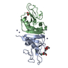
| ||||||||
|---|---|---|---|---|---|---|---|---|---|
| 1 |
| ||||||||
| 2 | 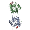
| ||||||||
| 3 | 
| ||||||||
| Unit cell |
| ||||||||
| Noncrystallographic symmetry (NCS) | NCS oper: (Code: given Matrix: (0.1615, -0.0535, -0.9854), Vector: |
-Components
-Protein , 1 types, 2 molecules AB
| #1: Protein | Mass: 14042.505 Da / Num. of mol.: 2 Source method: isolated from a genetically manipulated source Source: (gene. exp.) Polyandrocarpa misakiensis (invertebrata)Description: SYNTHETIC GENE / Plasmid: PRSETA-LEC Cellular location (production host): CYTOPLASM AS INCLUSION BODIES Gene (production host): TC14-1 / Production host:  |
|---|
-Non-polymers , 5 types, 296 molecules 


| #2: Chemical | ChemComp-CA /  Mass: 40.078 Da / Num. of mol.: 4 / Source method: obtained synthetically / Formula: Ca Mass: 40.078 Da / Num. of mol.: 4 / Source method: obtained synthetically / Formula: Ca#3: Chemical | ChemComp-ZN /  Mass: 65.409 Da / Num. of mol.: 7 / Source method: obtained synthetically / Formula: Zn Mass: 65.409 Da / Num. of mol.: 7 / Source method: obtained synthetically / Formula: Zn#4: Chemical | ChemComp-ACT /  Mass: 59.044 Da / Num. of mol.: 4 / Source method: obtained synthetically / Formula: C2H3O2 Mass: 59.044 Da / Num. of mol.: 4 / Source method: obtained synthetically / Formula: C2H3O2#5: Chemical |  Mass: 92.094 Da / Num. of mol.: 2 / Source method: obtained synthetically / Formula: C3H8O3 Mass: 92.094 Da / Num. of mol.: 2 / Source method: obtained synthetically / Formula: C3H8O3#6: Water | ChemComp-HOH / | Mass: 18.015 Da / Num. of mol.: 279 / Source method: isolated from a natural source / Formula: H2O |
|---|
-Details
| Has protein modification | Y |
|---|
-Experimental details
-Experiment
| Experiment | Method: X-RAY DIFFRACTION / Number of used crystals: 2 |
|---|
- Sample preparation
| Crystal | Density Matthews: 2.8 Å3/Da / Density % sol: 48 % | ||||||||||||||||||||||||||||||||||||||||||||||||||||||
|---|---|---|---|---|---|---|---|---|---|---|---|---|---|---|---|---|---|---|---|---|---|---|---|---|---|---|---|---|---|---|---|---|---|---|---|---|---|---|---|---|---|---|---|---|---|---|---|---|---|---|---|---|---|---|---|
| Crystal grow | pH: 6.5 Details: PROTEIN WAS CRYSTALLIZED FROM 7% PEG 8000, 0.2 M ZN(ACETATE) AND 100 MM NA( CACODYLATE), PH 6.5 | ||||||||||||||||||||||||||||||||||||||||||||||||||||||
| Crystal | *PLUS | ||||||||||||||||||||||||||||||||||||||||||||||||||||||
| Crystal grow | *PLUS Temperature: 17 ℃ / pH: 7.2 / Method: vapor diffusion, hanging drop | ||||||||||||||||||||||||||||||||||||||||||||||||||||||
| Components of the solutions | *PLUS
|
-Data collection
| Diffraction | Mean temperature: 100 K |
|---|---|
| Diffraction source | Source: ROTATING ANODE / Type: ELLIOTT GX-13 / Wavelength: 1.5418 |
| Detector | Type: MARRESEARCH / Detector: IMAGE PLATE / Date: Apr 15, 1998 / Details: MIRRORS |
| Radiation | Monochromator: MIRRORS / Protocol: SINGLE WAVELENGTH / Monochromatic (M) / Laue (L): M / Scattering type: x-ray |
| Radiation wavelength | Wavelength: 1.5418 Å / Relative weight: 1 |
| Reflection | Resolution: 2→52.7 Å / Num. obs: 20632 / % possible obs: 95.9 % / Redundancy: 6.6 % / Biso Wilson estimate: 17.63 Å2 / Rmerge(I) obs: 0.068 / Rsym value: 0.068 / Net I/σ(I): 8.3 |
| Reflection shell | Resolution: 2→2.05 Å / Redundancy: 5.6 % / Rmerge(I) obs: 0.185 / Mean I/σ(I) obs: 4 / Rsym value: 0.185 / % possible all: 87.9 |
| Reflection | *PLUS Num. measured all: 115502 |
| Reflection shell | *PLUS % possible obs: 87.9 % |
- Processing
| Software |
| ||||||||||||||||||||||||||||||||||||||||||||||||||||||||||||||||||||||||||||||||||||
|---|---|---|---|---|---|---|---|---|---|---|---|---|---|---|---|---|---|---|---|---|---|---|---|---|---|---|---|---|---|---|---|---|---|---|---|---|---|---|---|---|---|---|---|---|---|---|---|---|---|---|---|---|---|---|---|---|---|---|---|---|---|---|---|---|---|---|---|---|---|---|---|---|---|---|---|---|---|---|---|---|---|---|---|---|---|
| Refinement | Method to determine structure: SIRAS / Resolution: 2→35 Å / SU B: 3.8 / SU ML: 0.11 / Cross valid method: THROUGHOUT / σ(F): 0 / ESU R: 0.2 / ESU R Free: 0.17 Details: ELECTRON DENSITY FOR RESIDUE MET A 1 AND MET B 1 IS PRESENT BUT MODELLING WITH ACCEPTABLE GEOMETRY WAS NOT POSSIBLE. A CHAIN OF 5 VERY HIGH ELECTRON DENSITY SPHERES ADJACENT AND ORTHOGONAL ...Details: ELECTRON DENSITY FOR RESIDUE MET A 1 AND MET B 1 IS PRESENT BUT MODELLING WITH ACCEPTABLE GEOMETRY WAS NOT POSSIBLE. A CHAIN OF 5 VERY HIGH ELECTRON DENSITY SPHERES ADJACENT AND ORTHOGONAL TO THE SIDE-CHAIN OF ASP A 2 AND ASP B 2 WAS MODELLED AS ZN-HOH-ZN-HOH-ZN, BUT THE RESOLUTION IS NOT HIGH ENOUGH FOR UNAMBIGUOUS CHARACTERISATION.
| ||||||||||||||||||||||||||||||||||||||||||||||||||||||||||||||||||||||||||||||||||||
| Displacement parameters | Biso mean: 20 Å2 | ||||||||||||||||||||||||||||||||||||||||||||||||||||||||||||||||||||||||||||||||||||
| Refinement step | Cycle: LAST / Resolution: 2→35 Å
| ||||||||||||||||||||||||||||||||||||||||||||||||||||||||||||||||||||||||||||||||||||
| Refine LS restraints |
| ||||||||||||||||||||||||||||||||||||||||||||||||||||||||||||||||||||||||||||||||||||
| Software | *PLUS Name: REFMAC / Classification: refinement | ||||||||||||||||||||||||||||||||||||||||||||||||||||||||||||||||||||||||||||||||||||
| Refinement | *PLUS Highest resolution: 2 Å / σ(F): 0 / Rfactor obs: 0.206 / Rfactor Rfree: 0.25 | ||||||||||||||||||||||||||||||||||||||||||||||||||||||||||||||||||||||||||||||||||||
| Solvent computation | *PLUS | ||||||||||||||||||||||||||||||||||||||||||||||||||||||||||||||||||||||||||||||||||||
| Displacement parameters | *PLUS Biso mean: 20 Å2 | ||||||||||||||||||||||||||||||||||||||||||||||||||||||||||||||||||||||||||||||||||||
| Refine LS restraints | *PLUS
|
