 Movie
Movie Controller


+ Open data

- Basic information
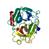
| Entry | Database: PDB / ID: 1b0f | |||||||||
|---|---|---|---|---|---|---|---|---|---|---|
| Title | CRYSTAL STRUCTURE OF HUMAN NEUTROPHIL ELASTASE WITH MDL 101, 146 | |||||||||
 Components Components | PROTEIN (ELASTASE) | |||||||||
 Keywords Keywords | HYDROLASE / SERINE PROTEASE / FLUOROETHYL KETONES | |||||||||
| Function / homology |  Function and homology information Function and homology informationleukocyte elastase / biosynthetic process of antibacterial peptides active against Gram-negative bacteria / Expression of NOTCH2NL genes / acute inflammatory response to antigenic stimulus / neutrophil-mediated killing of fungus / negative regulation of chemotaxis / positive regulation of leukocyte tethering or rolling / leukocyte migration involved in inflammatory response / response to yeast / negative regulation of chemokine production ...leukocyte elastase / biosynthetic process of antibacterial peptides active against Gram-negative bacteria / Expression of NOTCH2NL genes / acute inflammatory response to antigenic stimulus / neutrophil-mediated killing of fungus / negative regulation of chemotaxis / positive regulation of leukocyte tethering or rolling / leukocyte migration involved in inflammatory response / response to yeast / negative regulation of chemokine production / negative regulation of interleukin-8 production / Antimicrobial peptides / neutrophil-mediated killing of gram-negative bacterium / Activation of Matrix Metalloproteinases / positive regulation of MAP kinase activity / cytokine binding / Collagen degradation / extracellular matrix disassembly / Pyroptosis / pyroptotic inflammatory response / response to UV / phagocytosis / Degradation of the extracellular matrix / phagocytic vesicle / transcription repressor complex / secretory granule / positive regulation of smooth muscle cell proliferation / Regulation of Complement cascade / positive regulation of interleukin-8 production / protein catabolic process / specific granule lumen / negative regulation of inflammatory response / positive regulation of immune response / intracellular calcium ion homeostasis / azurophil granule lumen / transcription corepressor activity / peptidase activity / heparin binding / extracellular matrix / protease binding / endopeptidase activity / response to lipopolysaccharide / defense response to bacterium / serine-type endopeptidase activity / Neutrophil degranulation / cell surface / negative regulation of transcription by RNA polymerase II / Golgi apparatus / proteolysis / : / extracellular exosome / extracellular region / cytoplasm / cytosol Similarity search - Function | |||||||||
| Biological species |  Homo sapiens (human) Homo sapiens (human) | |||||||||
| Method |  X-RAY DIFFRACTION / MOLECULAR REPLACEMENT / Resolution: 3 Å X-RAY DIFFRACTION / MOLECULAR REPLACEMENT / Resolution: 3 Å | |||||||||
 Authors Authors | Schreuder, H.A. / Metz, W.A. / Peet, N.P. / Pelton, J.T. / Tardif, C. | |||||||||
 Citation Citation | Journal: J.Med.Chem. / Year: 1998 Title: Inhibition of human neutrophil elastase. 4. Design, synthesis, X-ray crystallographic analysis, and structure-activity relationships for a series of P2-modified, orally active peptidyl pentafluoroethyl ketones. Authors: Cregge, R.J. / Durham, S.L. / Farr, R.A. / Gallion, S.L. / Hare, C.M. / Hoffman, R.V. / Janusz, M.J. / Kim, H.O. / Koehl, J.R. / Mehdi, S. / Metz, W.A. / Peet, N.P. / Pelton, J.T. / ...Authors: Cregge, R.J. / Durham, S.L. / Farr, R.A. / Gallion, S.L. / Hare, C.M. / Hoffman, R.V. / Janusz, M.J. / Kim, H.O. / Koehl, J.R. / Mehdi, S. / Metz, W.A. / Peet, N.P. / Pelton, J.T. / Schreuder, H.A. / Sunder, S. / Tardif, C. | |||||||||
| History |
|
- Structure visualization
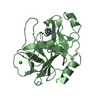
| Structure viewer | Molecule: MolmilJmol/JSmol |
|---|
- Downloads & links
-Download
| PDBx/mmCIF format | 1b0f.cif.gz | 58.5 KB | Display | PDBx/mmCIF format |
|---|---|---|---|---|
| PDB format | pdb1b0f.ent.gz | 41.5 KB | Display | PDB format |
| PDBx/mmJSON format | 1b0f.json.gz | Tree view | PDBx/mmJSON format | |
| Others |  Other downloads Other downloads |
-Validation report
| Arichive directory | https://data.pdbj.org/pub/pdb/validation_reports/b0/1b0fftp://data.pdbj.org/pub/pdb/validation_reports/b0/1b0f | HTTPS FTP |
|---|
-Related structure data
| Related structure data | 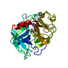 1b0eC  1hneS S: Starting model for refinement C: citing same article ( |
|---|---|
| Similar structure data |


-Links
 PDBj
PDBj


- Assembly
| Deposited unit | 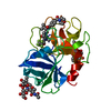
| ||||||||
|---|---|---|---|---|---|---|---|---|---|
| 1 |
| ||||||||
| Unit cell |
|
-Components
| #1: Protein | Mass: 23233.850 Da / Num. of mol.: 1 / Source method: isolated from a natural source / Source: (natural) Homo sapiens (human) / Cell: NEUTROPHIL / References: UniProt: P08246, leukocyte elastase | ||||||
|---|---|---|---|---|---|---|---|
| #2: Polysaccharide | Source method: isolated from a genetically manipulated source #3: Chemical | ChemComp-SEI / | 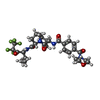  Mass: 632.619 Da / Num. of mol.: 1 / Source method: obtained synthetically / Formula: C29H37F5N4O6 Mass: 632.619 Da / Num. of mol.: 1 / Source method: obtained synthetically / Formula: C29H37F5N4O6#4: Water | ChemComp-HOH / |  Mass: 18.015 Da / Num. of mol.: 34 / Source method: isolated from a natural source / Formula: H2O Mass: 18.015 Da / Num. of mol.: 34 / Source method: isolated from a natural source / Formula: H2OHas protein modification | Y | |
-Experimental details
-Experiment
| Experiment | Method: X-RAY DIFFRACTION / Number of used crystals: 1 |
|---|
- Sample preparation
| Crystal | Density Matthews: 3.2 Å3/Da / Density % sol: 63.31 % | ||||||||||||||||||||||||||||||||||||
|---|---|---|---|---|---|---|---|---|---|---|---|---|---|---|---|---|---|---|---|---|---|---|---|---|---|---|---|---|---|---|---|---|---|---|---|---|---|
| Crystal grow | pH: 5 / Details: pH 5.00 | ||||||||||||||||||||||||||||||||||||
| Crystal grow | *PLUS Temperature: 4 ℃ / Method: vapor diffusion, hanging drop / pH: 3.8 | ||||||||||||||||||||||||||||||||||||
| Components of the solutions | *PLUS
|
-Data collection
| Diffraction | Mean temperature: 287 K |
|---|---|
| Diffraction source | Source: ROTATING ANODE / Type: SIEMENS / Wavelength: 1.5418 |
| Detector | Type: SIEMENS / Detector: AREA DETECTOR / Date: Oct 19, 1993 / Details: COLLIMATOR |
| Radiation | Monochromator: GRAPHITE / Protocol: SINGLE WAVELENGTH / Monochromatic (M) / Laue (L): M / Scattering type: x-ray |
| Radiation wavelength | Wavelength: 1.5418 Å / Relative weight: 1 |
| Reflection | Resolution: 3→20 Å / Num. obs: 6729 / % possible obs: 99.1 % / Redundancy: 7.2 % / Rsym value: 0.099 |
| Reflection shell | Resolution: 3→3.1 Å / Redundancy: 5.2 % / Rsym value: 0.371 / % possible all: 95.9 |
| Reflection | *PLUS Highest resolution: 3 Å / Lowest resolution: 20 Å / Redundancy: 7.2 % / Num. measured all: 48509 / Rmerge(I) obs: 0.099 |
| Reflection shell | *PLUS Highest resolution: 3 Å / Lowest resolution: 3.1 Å / Redundancy: 5.2 % |
- Processing
| Software |
| ||||||||||||||||||||||||||||||||||||||||||||||||||||||||||||||||||||||||||||||||
|---|---|---|---|---|---|---|---|---|---|---|---|---|---|---|---|---|---|---|---|---|---|---|---|---|---|---|---|---|---|---|---|---|---|---|---|---|---|---|---|---|---|---|---|---|---|---|---|---|---|---|---|---|---|---|---|---|---|---|---|---|---|---|---|---|---|---|---|---|---|---|---|---|---|---|---|---|---|---|---|---|---|
| Refinement | Method to determine structure: MOLECULAR REPLACEMENT Starting model: PDB ENTRY 1HNE Resolution: 3→8 Å / Data cutoff high absF: 100000 / Data cutoff low absF: 0.1 / Isotropic thermal model: RESTRAINED / σ(F): 2
| ||||||||||||||||||||||||||||||||||||||||||||||||||||||||||||||||||||||||||||||||
| Displacement parameters | Biso mean: 21 Å2 | ||||||||||||||||||||||||||||||||||||||||||||||||||||||||||||||||||||||||||||||||
| Refine analyze | Luzzati coordinate error obs: 0.25 Å | ||||||||||||||||||||||||||||||||||||||||||||||||||||||||||||||||||||||||||||||||
| Refinement step | Cycle: LAST / Resolution: 3→8 Å
| ||||||||||||||||||||||||||||||||||||||||||||||||||||||||||||||||||||||||||||||||
| Refine LS restraints |
| ||||||||||||||||||||||||||||||||||||||||||||||||||||||||||||||||||||||||||||||||
| LS refinement shell | Resolution: 3→3.13 Å / Total num. of bins used: 8
| ||||||||||||||||||||||||||||||||||||||||||||||||||||||||||||||||||||||||||||||||
| Xplor file |
| ||||||||||||||||||||||||||||||||||||||||||||||||||||||||||||||||||||||||||||||||
| Software | *PLUS Name: X-PLOR / Version: 3.1 / Classification: refinement | ||||||||||||||||||||||||||||||||||||||||||||||||||||||||||||||||||||||||||||||||
| Refinement | *PLUS Rfactor obs: 0.16 / Rfactor Rwork: 0.16 | ||||||||||||||||||||||||||||||||||||||||||||||||||||||||||||||||||||||||||||||||
| Solvent computation | *PLUS | ||||||||||||||||||||||||||||||||||||||||||||||||||||||||||||||||||||||||||||||||
| Displacement parameters | *PLUS | ||||||||||||||||||||||||||||||||||||||||||||||||||||||||||||||||||||||||||||||||
| Refine LS restraints | *PLUS
| ||||||||||||||||||||||||||||||||||||||||||||||||||||||||||||||||||||||||||||||||
| LS refinement shell | *PLUS Rfactor Rwork: 0.206 |
