 Movie
Movie Controller


+ Open data

- Basic information
| Entry | Database: PDB / ID: 1axk | ||||||
|---|---|---|---|---|---|---|---|
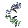
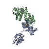
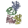
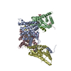
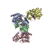
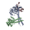
| Title | ENGINEERED BACILLUS BIFUNCTIONAL ENZYME GLUXYN-1 | ||||||
 Components Components | GLUXYN-1 | ||||||
 Keywords Keywords | FUSION PROTEIN / GLUXYN-1 / BIFUNCTIONAL / 1 / 4-BETA-XYLANASE / 3-1 / 4-BETA-GLUCANASE / HYBRID ENZYME | ||||||
| Function / homology |  Function and homology information Function and homology informationlicheninase / licheninase activity / endo-1,4-beta-xylanase / endo-1,4-beta-xylanase activity / xylan catabolic process / carbohydrate metabolic process Similarity search - Function | ||||||
| Biological species |  | ||||||
| Method |  X-RAY DIFFRACTION / SYNCHROTRON / MOLECULAR REPLACEMENT / Resolution: 2.1 Å X-RAY DIFFRACTION / SYNCHROTRON / MOLECULAR REPLACEMENT / Resolution: 2.1 Å | ||||||
 Authors Authors | Ay, J. / Heinemann, U. | ||||||
 Citation Citation | Journal: Proc.Natl.Acad.Sci.USA / Year: 1998 Title: Structure and function of the Bacillus hybrid enzyme GluXyn-1: native-like jellyroll fold preserved after insertion of autonomous globular domain. Authors: Ay, J. / Gotz, F. / Borriss, R. / Heinemann, U. | ||||||
| History |
|
- Structure visualization
| Structure viewer | Molecule: MolmilJmol/JSmol |
|---|
- Downloads & links
-Download
| PDBx/mmCIF format | 1axk.cif.gz | 169.6 KB | Display | PDBx/mmCIF format |
|---|---|---|---|---|
| PDB format | pdb1axk.ent.gz | 133.8 KB | Display | PDB format |
| PDBx/mmJSON format | 1axk.json.gz | Tree view | PDBx/mmJSON format | |
| Others |  Other downloads Other downloads |
-Validation report
| Arichive directory | https://data.pdbj.org/pub/pdb/validation_reports/ax/1axkftp://data.pdbj.org/pub/pdb/validation_reports/ax/1axk | HTTPS FTP |
|---|
-Related structure data


-Links
 PDBj
PDBj

- Assembly
| Deposited unit | 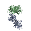
| ||||||||||||||||
|---|---|---|---|---|---|---|---|---|---|---|---|---|---|---|---|---|---|
| 1 |
| ||||||||||||||||
| Unit cell |
| ||||||||||||||||
| Noncrystallographic symmetry (NCS) | NCS oper:
|
-Components
| #1: Protein | Mass: 43812.676 Da / Num. of mol.: 2 Fragment: FUSION OF 1,3-1,4-BETA-GLUCANASE DOMAIN AND 1,4-BETA-XYLANASE DOMAIN Source method: isolated from a genetically manipulated source Details: ACTIVE AS BOTH A 1,3-1,4-BETA-GLUCANASE AND A 1,4-BETA-XYLANASE Source: (gene. exp.) References: UniProt: P23904, UniProt: P18429, licheninase, endo-1,4-beta-xylanase #2: Chemical |   Mass: 40.078 Da / Num. of mol.: 2 / Source method: obtained synthetically / Formula: Ca Mass: 40.078 Da / Num. of mol.: 2 / Source method: obtained synthetically / Formula: Ca#3: Water | ChemComp-HOH / |  Mass: 18.015 Da / Num. of mol.: 312 / Source method: isolated from a natural source / Formula: H2O Mass: 18.015 Da / Num. of mol.: 312 / Source method: isolated from a natural source / Formula: H2OHas protein modification | Y | |
|---|
-Experimental details
-Experiment
| Experiment | Method: X-RAY DIFFRACTION / Number of used crystals: 1 |
|---|
- Sample preparation
| Crystal | Density Matthews: 2.6 Å3/Da / Density % sol: 46.4 % | ||||||||||||||||||||||||||||||||||||
|---|---|---|---|---|---|---|---|---|---|---|---|---|---|---|---|---|---|---|---|---|---|---|---|---|---|---|---|---|---|---|---|---|---|---|---|---|---|
| Crystal grow | pH: 8.5 / Details: pH 8.5 | ||||||||||||||||||||||||||||||||||||
| Crystal grow | *PLUS pH: 8.7 / Method: vapor diffusion, hanging drop | ||||||||||||||||||||||||||||||||||||
| Components of the solutions | *PLUS
|
-Data collection
| Diffraction | Mean temperature: 123 K |
|---|---|
| Diffraction source | Source: SYNCHROTRON / Site: ESRF  / Beamline: BM14 / Wavelength: 1.008 / Beamline: BM14 / Wavelength: 1.008 |
| Detector | Type: MARRESEARCH / Detector: IMAGE PLATE / Date: Aug 1, 1996 |
| Radiation | Monochromatic (M) / Laue (L): M / Scattering type: x-ray |
| Radiation wavelength | Wavelength: 1.008 Å / Relative weight: 1 |
| Reflection | Resolution: 2.1→20 Å / Num. obs: 48499 / % possible obs: 91.3 % / Observed criterion σ(I): 0 / Redundancy: 3.4 % / Biso Wilson estimate: 23.98 Å2 / Rmerge(I) obs: 0.058 / Rsym value: 0.058 / Net I/σ(I): 11.6 |
| Reflection shell | Resolution: 2.1→2.21 Å / Redundancy: 3.2 % / Rmerge(I) obs: 0.23 / Mean I/σ(I) obs: 3.3 / Rsym value: 0.23 / % possible all: 80 |
| Reflection | *PLUS Num. measured all: 165157 |
| Reflection shell | *PLUS Lowest resolution: 2.17 Å / % possible obs: 80 % |
- Processing
| Software |
| ||||||||||||||||||||||||||||||||||||||||||||||||||||||||||||||||||||||||||||||||||||
|---|---|---|---|---|---|---|---|---|---|---|---|---|---|---|---|---|---|---|---|---|---|---|---|---|---|---|---|---|---|---|---|---|---|---|---|---|---|---|---|---|---|---|---|---|---|---|---|---|---|---|---|---|---|---|---|---|---|---|---|---|---|---|---|---|---|---|---|---|---|---|---|---|---|---|---|---|---|---|---|---|---|---|---|---|---|
| Refinement | Method to determine structure: MOLECULAR REPLACEMENT Starting model: CIRCULARLY PERMUTED 1,3-1,4-ENDO-BETA-GLUCANASE CPMAC57 (PDB ENTRY 1CPN) AND BACILLUS CIRCULANS 1,3-ENDO-BETA-XYLANASE (PDB ENTRY 1BCX). Resolution: 2.1→19.96 Å / Cross valid method: THROUGHOUT / σ(F): 0
| ||||||||||||||||||||||||||||||||||||||||||||||||||||||||||||||||||||||||||||||||||||
| Displacement parameters | Biso mean: 21.7 Å2
| ||||||||||||||||||||||||||||||||||||||||||||||||||||||||||||||||||||||||||||||||||||
| Refine analyze | Luzzati coordinate error obs: 0.3 Å | ||||||||||||||||||||||||||||||||||||||||||||||||||||||||||||||||||||||||||||||||||||
| Refinement step | Cycle: LAST / Resolution: 2.1→19.96 Å
| ||||||||||||||||||||||||||||||||||||||||||||||||||||||||||||||||||||||||||||||||||||
| Refine LS restraints |
|
