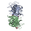
 Movie
Movie Controller


+ Open data

- Basic information
| Entry | Database: PDB / ID: 1avb | |||||||||
|---|---|---|---|---|---|---|---|---|---|---|
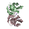
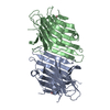
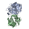
| Title | ARCELIN-1 FROM PHASEOLUS VULGARIS L | |||||||||
 Components Components | ARCELIN-1 | |||||||||
 Keywords Keywords | LECTIN / LECTIN-LIKE GLYCOPROTEIN / PLANT DEFENSE / INSECTICIDAL ACTIVITY | |||||||||
| Function / homology |  Function and homology information Function and homology informationnutrient reservoir activity / defense response / toxin activity / carbohydrate binding Similarity search - Function | |||||||||
| Biological species |  Phaseolus vulgaris (common bean) Phaseolus vulgaris (common bean) | |||||||||
| Method |  X-RAY DIFFRACTION / SYNCHROTRON / MIR, DENSITY MODIFICATION, MOLECULAR REPLACEMENT / Resolution: 1.9 Å X-RAY DIFFRACTION / SYNCHROTRON / MIR, DENSITY MODIFICATION, MOLECULAR REPLACEMENT / Resolution: 1.9 Å | |||||||||
 Authors Authors | Mourey, L. / Pedelacq, J.D. / Fabre, C. / Rouge, P. / Samama, J.P. | |||||||||
 Citation Citation | Journal: J.Biol.Chem. / Year: 1998 Title: Crystal structure of the arcelin-1 dimer from Phaseolus vulgaris at 1.9-A resolution. Authors: Mourey, L. / Pedelacq, J.D. / Birck, C. / Fabre, C. / Rouge, P. / Samama, J.P. #1: Journal: Proteins / Year: 1997Title: Small-Angle X-Ray Scattering and Crystallographic Studies of Arcelin-1: An Insecticidal Lectin-Like Glycoprotein from Phaseolus Vulgaris L Authors: Mourey, L. / Pedelacq, J.D. / Fabre, C. / Causse, H. / Rouge, P. / Samama, J.P. | |||||||||
| History |
|
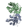
- Structure visualization
| Structure viewer | Molecule: MolmilJmol/JSmol |
|---|
- Downloads & links
-Download
| PDBx/mmCIF format | 1avb.cif.gz | 105.6 KB | Display | PDBx/mmCIF format |
|---|---|---|---|---|
| PDB format | pdb1avb.ent.gz | 81.9 KB | Display | PDB format |
| PDBx/mmJSON format | 1avb.json.gz | Tree view | PDBx/mmJSON format | |
| Others |  Other downloads Other downloads |
-Validation report
| Arichive directory | https://data.pdbj.org/pub/pdb/validation_reports/av/1avbftp://data.pdbj.org/pub/pdb/validation_reports/av/1avb | HTTPS FTP |
|---|
-Related structure data
| Related structure data | 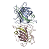 1loeS S: Starting model for refinement |
|---|---|
| Similar structure data |


-Links
 PDBj
PDBj

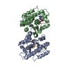
- Assembly
| Deposited unit | 
| ||||||||
|---|---|---|---|---|---|---|---|---|---|
| 1 |
| ||||||||
| Unit cell |
| ||||||||
| Noncrystallographic symmetry (NCS) | NCS oper: (Code: given Matrix: (-0.999998, -0.001916, 0.000892), Vector: |
-Components
| #1: Protein | Mass: 24984.553 Da / Num. of mol.: 2 / Source method: isolated from a natural source / Source: (natural) Phaseolus vulgaris (common bean) / Organ: SEED / Strain: CV. RAZ-2 / References: UniProt: P19329#2: Polysaccharide | Source method: isolated from a genetically manipulated source #3: Sugar | ChemComp-NAG /   Type: D-saccharide, beta linking / Mass: 221.208 Da / Num. of mol.: 4 Type: D-saccharide, beta linking / Mass: 221.208 Da / Num. of mol.: 4Source method: isolated from a genetically manipulated source Formula: C8H15NO6 #4: Chemical |   Mass: 96.063 Da / Num. of mol.: 2 / Source method: obtained synthetically / Formula: SO4 Mass: 96.063 Da / Num. of mol.: 2 / Source method: obtained synthetically / Formula: SO4#5: Water | ChemComp-HOH / |  Mass: 18.015 Da / Num. of mol.: 230 / Source method: isolated from a natural source / Formula: H2O Mass: 18.015 Da / Num. of mol.: 230 / Source method: isolated from a natural source / Formula: H2OHas protein modification | Y | |
|---|
-Experimental details
-Experiment
| Experiment | Method: X-RAY DIFFRACTION / Number of used crystals: 1 |
|---|
- Sample preparation
| Crystal | Density Matthews: 2.2 Å3/Da / Density % sol: 44 % | ||||||||||||||||||||||||||||||
|---|---|---|---|---|---|---|---|---|---|---|---|---|---|---|---|---|---|---|---|---|---|---|---|---|---|---|---|---|---|---|---|
| Crystal grow | pH: 4.8 Details: PROTEIN WAS CRYSTALLIZED FROM 25% PEG400, 0.3 M AMMONIUM SULFATE IN 0.1 M MES BUFFER PH 4.8-4.9 PH range: 4.8-4.9 | ||||||||||||||||||||||||||||||
| Crystal | *PLUS | ||||||||||||||||||||||||||||||
| Crystal grow | *PLUS Temperature: 4 ℃ / Method: vapor diffusion, hanging dropDetails: Mourey, L., (1997) Proteins: Struct.,Funct., Genet., 29, 433. | ||||||||||||||||||||||||||||||
| Components of the solutions | *PLUS
|
-Data collection
| Diffraction | Mean temperature: 277 K |
|---|---|
| Diffraction source | Source: SYNCHROTRON / Site: LURE  / Beamline: DW32 / Wavelength: 0.938 / Beamline: DW32 / Wavelength: 0.938 |
| Detector | Type: MARRESEARCH / Detector: IMAGE PLATE / Date: Oct 1, 1995 |
| Radiation | Monochromatic (M) / Laue (L): M / Scattering type: x-ray |
| Radiation wavelength | Wavelength: 0.938 Å / Relative weight: 1 |
| Reflection | Resolution: 1.9→33.7 Å / Num. obs: 41612 / % possible obs: 97.7 % / Redundancy: 3 % / Biso Wilson estimate: 15.6 Å2 / Rmerge(I) obs: 0.064 / Net I/σ(I): 11.9 |
| Reflection shell | Resolution: 1.9→1.95 Å / Redundancy: 2.6 % / Rmerge(I) obs: 0.157 / Mean I/σ(I) obs: 7.5 / % possible all: 93.9 |
- Processing
| Software |
| ||||||||||||||||||||||||||||||||||||||||||||||||||||||||||||||||||||||||||||||||
|---|---|---|---|---|---|---|---|---|---|---|---|---|---|---|---|---|---|---|---|---|---|---|---|---|---|---|---|---|---|---|---|---|---|---|---|---|---|---|---|---|---|---|---|---|---|---|---|---|---|---|---|---|---|---|---|---|---|---|---|---|---|---|---|---|---|---|---|---|---|---|---|---|---|---|---|---|---|---|---|---|---|
| Refinement | Method to determine structure: MIR, DENSITY MODIFICATION, MOLECULAR REPLACEMENT Starting model: PDB ENTRY 1LOE Resolution: 1.9→33.71 Å / Rfactor Rfree error: 0.005 / Data cutoff high absF: 100000 / Data cutoff low absF: 0.1 / Cross valid method: THROUGHOUT / σ(F): 2 Details: NCS CONSTRAINTS WERE APPLIED DURING REFINEMENT TO 2.5 ANGSTROMS RESOLUTION. THEY WERE LATER RELEASED AFTER RESOLUTION WAS EXTENDED TO 1.9 ANGSTROMS.
| ||||||||||||||||||||||||||||||||||||||||||||||||||||||||||||||||||||||||||||||||
| Displacement parameters | Biso mean: 22.2 Å2 | ||||||||||||||||||||||||||||||||||||||||||||||||||||||||||||||||||||||||||||||||
| Refine analyze |
| ||||||||||||||||||||||||||||||||||||||||||||||||||||||||||||||||||||||||||||||||
| Refinement step | Cycle: LAST / Resolution: 1.9→33.71 Å
| ||||||||||||||||||||||||||||||||||||||||||||||||||||||||||||||||||||||||||||||||
| Refine LS restraints |
| ||||||||||||||||||||||||||||||||||||||||||||||||||||||||||||||||||||||||||||||||
| Refine LS restraints NCS | NCS model details: UNRESTRAINED | ||||||||||||||||||||||||||||||||||||||||||||||||||||||||||||||||||||||||||||||||
| LS refinement shell | Resolution: 1.9→1.93 Å / Rfactor Rfree error: 0.029 / Total num. of bins used: 15
| ||||||||||||||||||||||||||||||||||||||||||||||||||||||||||||||||||||||||||||||||
| Xplor file |
| ||||||||||||||||||||||||||||||||||||||||||||||||||||||||||||||||||||||||||||||||
| Software | *PLUS Name: X-PLOR / Version: 3.1 / Classification: refinement | ||||||||||||||||||||||||||||||||||||||||||||||||||||||||||||||||||||||||||||||||
| Refine LS restraints | *PLUS
|
