- PDB-3cwq: Crystal structure of chromosome partitioning protein (ParA) in co... -
+
Open data
ID or keywords:
Loading...
-
Basic information
Entry
Database: PDB / ID: 3cwq
Title
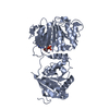
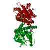
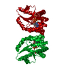
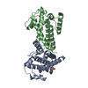
Crystal structure of chromosome partitioning protein (ParA) in complex with ADP from Synechocystis sp. Northeast Structural Genomics Consortium Target SgR89
Components
ParA family chromosome partitioning protein
Keywords
STRUCTURAL GENOMICS / UNKNOWN FUNCTION / alpha-beta protein / PSI-2 / Protein Structure Initiative / Northeast Structural Genomics Consortium / NESG
Function / homology
Function and homology information
cytoplasmic side of plasma membrane / ATP hydrolysis activity / ATP binding / cytosol Similarity search - Function
Mass: 18.015 Da / Num. of mol.: 39 / Source method: isolated from a natural source / Formula: H2O
Has protein modification
Y
-
Experimental details
-
Experiment
Experiment
Method: X-RAY DIFFRACTION / Number of used crystals: 1
-
Sample preparation
Crystal
Density Matthews: 2.73 Å3/Da / Density % sol: 54.94 %
Crystal grow
Temperature: 277 K / Method: microbatch under oil / pH: 7.5 Details: Protein solution: 10 mM Tris-HCl pH 7.5, 100 Sodium chloride, 5 mM DTT. Resevoir solution: 25% Ethylene glycol, MICROBATCH UNDER OIL, temperature 277K
Resolution: 2.47→2.54 Å / Redundancy: 6.3 % / Rmerge(I) obs: 0.22 / Mean I/σ(I) obs: 6.96 / Rsym value: 0.155 / % possible all: 90.7
-
Processing
Software
Name
Version
Classification
REFMAC
5.2.0003
refinement
ADSC
Quantum
datacollection
DENZO
datareduction
SCALEPACK
datascaling
SnB
phasing
SOLVE
phasing
RESOLVE
phasing
Refinement
Method to determine structure: SAD / Resolution: 2.47→29.62 Å / Cor.coef. Fo:Fc: 0.933 / Cor.coef. Fo:Fc free: 0.889 / SU B: 25.339 / SU ML: 0.253 / TLS residual ADP flag: LIKELY RESIDUAL / Cross valid method: THROUGHOUT / ESU R: 0.563 / ESU R Free: 0.318 / Stereochemistry target values: MAXIMUM LIKELIHOOD Details: The Friedel pairs were used in phasing. Program CNS 1.2 has also been used in refinement.
Rfactor
Num. reflection
% reflection
Selection details
Rfree
0.28173
842
5.1 %
RANDOM
Rwork
0.22069
-
-
-
obs
0.22368
15781
100 %
-
Solvent computation
Ion probe radii: 0.8 Å / Shrinkage radii: 0.8 Å / VDW probe radii: 1.2 Å / Solvent model: BABINET MODEL WITH MASK
Displacement parameters
Biso mean: 44.621 Å2
Baniso -1
Baniso -2
Baniso -3
1-
-4.83 Å2
0 Å2
0 Å2
2-
-
-7.08 Å2
0 Å2
3-
-
-
11.91 Å2
Refinement step
Cycle: LAST / Resolution: 2.47→29.62 Å
Protein
Nucleic acid
Ligand
Solvent
Total
Num. atoms
3074
0
54
39
3167
Refine LS restraints
Refine-ID
Type
Dev ideal
Dev ideal target
Number
X-RAY DIFFRACTION
r_bond_refined_d
0.031
0.022
3190
X-RAY DIFFRACTION
r_bond_other_d
X-RAY DIFFRACTION
r_angle_refined_deg
2.669
1.993
4338
X-RAY DIFFRACTION
r_angle_other_deg
X-RAY DIFFRACTION
r_dihedral_angle_1_deg
7.969
5
400
X-RAY DIFFRACTION
r_dihedral_angle_2_deg
40.472
24.426
122
X-RAY DIFFRACTION
r_dihedral_angle_3_deg
23.585
15
550
X-RAY DIFFRACTION
r_dihedral_angle_4_deg
16.947
15
18
X-RAY DIFFRACTION
r_chiral_restr
0.167
0.2
509
X-RAY DIFFRACTION
r_gen_planes_refined
0.009
0.02
2327
X-RAY DIFFRACTION
r_gen_planes_other
X-RAY DIFFRACTION
r_nbd_refined
0.274
0.2
1533
X-RAY DIFFRACTION
r_nbd_other
X-RAY DIFFRACTION
r_nbtor_refined
0.333
0.2
2177
X-RAY DIFFRACTION
r_nbtor_other
X-RAY DIFFRACTION
r_xyhbond_nbd_refined
0.212
0.2
117
X-RAY DIFFRACTION
r_xyhbond_nbd_other
X-RAY DIFFRACTION
r_metal_ion_refined
X-RAY DIFFRACTION
r_metal_ion_other
X-RAY DIFFRACTION
r_symmetry_vdw_refined
0.308
0.2
13
X-RAY DIFFRACTION
r_symmetry_vdw_other
X-RAY DIFFRACTION
r_symmetry_hbond_refined
0.232
0.2
2
X-RAY DIFFRACTION
r_symmetry_hbond_other
X-RAY DIFFRACTION
r_symmetry_metal_ion_refined
X-RAY DIFFRACTION
r_symmetry_metal_ion_other
X-RAY DIFFRACTION
r_mcbond_it
1.36
1.5
2080
X-RAY DIFFRACTION
r_mcbond_other
X-RAY DIFFRACTION
r_mcangle_it
2.089
2
3219
X-RAY DIFFRACTION
r_scbond_it
3.351
3
1306
X-RAY DIFFRACTION
r_scangle_it
4.931
4.5
1119
X-RAY DIFFRACTION
r_rigid_bond_restr
X-RAY DIFFRACTION
r_sphericity_free
X-RAY DIFFRACTION
r_sphericity_bonded
LS refinement shell
Resolution: 2.47→2.537 Å / Total num. of bins used: 20
Rfactor
Num. reflection
% reflection
Rfree
0.383
48
-
Rwork
0.274
788
-
obs
-
-
100 %
Refinement TLS params.
Method: refined / Origin x: 74.1905 Å / Origin y: 38.4644 Å / Origin z: 65.5746 Å
11
12
13
21
22
23
31
32
33
T
-0.0214 Å2
0.0064 Å2
-0.0135 Å2
-
0.0842 Å2
-0.032 Å2
-
-
-0.1771 Å2
L
1.6093 °2
1.244 °2
-0.4318 °2
-
4.6662 °2
-0.7383 °2
-
-
0.5751 °2
S
0.061 Å °
-0.1537 Å °
-0.0576 Å °
0.1562 Å °
-0.0591 Å °
-0.1534 Å °
-0.0106 Å °
0.0287 Å °
-0.0019 Å °
Refinement TLS group
ID
Refine-ID
Refine TLS-ID
Auth asym-ID
Auth seq-ID
1
X-RAY DIFFRACTION
1
B
302 - 320
2
X-RAY DIFFRACTION
1
A
302 - 322
3
X-RAY DIFFRACTION
1
B
301
4
X-RAY DIFFRACTION
1
A
301
5
X-RAY DIFFRACTION
1
B
1 - 201
6
X-RAY DIFFRACTION
1
A
1 - 206
+
About Yorodumi
-
News
-
Feb 9, 2022. New format data for meta-information of EMDB entries
New format data for meta-information of EMDB entries
Version 3 of the EMDB header file is now the official format.
The previous official version 1.9 will be removed from the archive.
In the structure databanks used in Yorodumi, some data are registered as the other names, "COVID-19 virus" and "2019-nCoV". Here are the details of the virus and the list of structure data.
Jan 31, 2019. EMDB accession codes are about to change! (news from PDBe EMDB page)
EMDB accession codes are about to change! (news from PDBe EMDB page)
The allocation of 4 digits for EMDB accession codes will soon come to an end. Whilst these codes will remain in use, new EMDB accession codes will include an additional digit and will expand incrementally as the available range of codes is exhausted. The current 4-digit format prefixed with “EMD-” (i.e. EMD-XXXX) will advance to a 5-digit format (i.e. EMD-XXXXX), and so on. It is currently estimated that the 4-digit codes will be depleted around Spring 2019, at which point the 5-digit format will come into force.
The EM Navigator/Yorodumi systems omit the EMD- prefix.
Related info.:Q: What is EMD? / ID/Accession-code notation in Yorodumi/EM Navigator
Yorodumi is a browser for structure data from EMDB, PDB, SASBDB, etc.
This page is also the successor to EM Navigator detail page, and also detail information page/front-end page for Omokage search.
The word "yorodu" (or yorozu) is an old Japanese word meaning "ten thousand". "mi" (miru) is to see.
Related info.:EMDB / PDB / SASBDB / Comparison of 3 databanks / Yorodumi Search / Aug 31, 2016. New EM Navigator & Yorodumi / Yorodumi Papers / Jmol/JSmol / Function and homology information / Changes in new EM Navigator and Yorodumi
 Movie
Movie Controller

 Yorodumi
 Open data
 Basic information
 Components
 Keywords
 Function and homology information
 X-RAY DIFFRACTION /
  Authors
 Citation
 Structure visualization
 Downloads & links
 Other downloads


 PDBj
PDBj Assembly
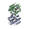

 Mass: 427.201 Da / Num. of mol.: 2 / Source method: obtained synthetically / Formula: C10H15N5O10P2 / Comment: ADP, energy-carrying molecule*YM
 Mass: 18.015 Da / Num. of mol.: 39 / Source method: isolated from a natural source / Formula: H2O
 Sample preparation
 / Beamline: X4A / Wavelength: 0.97898 Å
 Processing