 Movie
Movie Controller

[English] 日本語
 Yorodumi
- PDB-1h3m: Structure of 4-diphosphocytidyl-2C-methyl-D-erythritol synthetase -
+ Open data

- Basic information
| Entry | Database: PDB / ID: 1h3m | ||||||
|---|---|---|---|---|---|---|---|
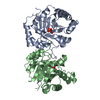
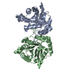
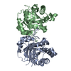
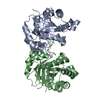
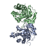
| Title | Structure of 4-diphosphocytidyl-2C-methyl-D-erythritol synthetase | ||||||
 Components Components | 2-C-METHYL-D-ERYTHRITOL 4-PHOSPHATE CYTIDYLYLTRANSFERASE | ||||||
 Keywords Keywords | TRANSFERASE / ISOPRENOID / SYNTHETASE | ||||||
| Function / homology |  Function and homology information Function and homology information2-C-methyl-D-erythritol 4-phosphate cytidylyltransferase / 2-C-methyl-D-erythritol 4-phosphate cytidylyltransferase activity / isopentenyl diphosphate biosynthetic process, methylerythritol 4-phosphate pathway / magnesium ion binding / identical protein binding / cytosol Similarity search - Function | ||||||
| Biological species |  | ||||||
| Method |  X-RAY DIFFRACTION / SYNCHROTRON / MOLECULAR REPLACEMENT / Resolution: 2.4 Å X-RAY DIFFRACTION / SYNCHROTRON / MOLECULAR REPLACEMENT / Resolution: 2.4 Å | ||||||
 Authors Authors | Kemp, L.E. / Bond, C.S. / Hunter, W.N. | ||||||
 Citation Citation | Journal: Acta Crystallogr.,Sect.D / Year: 2003 Title: Structure of a Tetragonal Crystal Form of Escherichia Coli 2-C-Methyl-D-Erythritol 4-Phosphate Cytidylyltransferase Authors: Kemp, L.E. / Bond, C.S. / Hunter, W.N. #1: Journal: Nat.Struct.Biol. / Year: 2001Title: Structure of Cdp-Me Synthetase in Mevalonate-Independent Isoprenoid Biosynthesis Authors: Richard, S.B. / Bowman, M.E. / Kwiatkowski, W. / Chow, C. / Lillo, A.M. / Cane, D.E. / Noel, J.P. | ||||||
| History |
| ||||||
| Remark 700 | SHEET THE SHEET STRUCTURE OF THIS MOLECULE IS BIFURCATED. IN ORDER TO REPRESENT THIS FEATURE IN ... SHEET THE SHEET STRUCTURE OF THIS MOLECULE IS BIFURCATED. IN ORDER TO REPRESENT THIS FEATURE IN THE SHEET RECORDS BELOW, TWO SHEETS ARE DEFINED. |
- Structure visualization
| Structure viewer | Molecule: MolmilJmol/JSmol |
|---|
- Downloads & links
-Download
| PDBx/mmCIF format | 1h3m.cif.gz | 97.7 KB | Display | PDBx/mmCIF format |
|---|---|---|---|---|
| PDB format | pdb1h3m.ent.gz | 74.7 KB | Display | PDB format |
| PDBx/mmJSON format | 1h3m.json.gz | Tree view | PDBx/mmJSON format | |
| Others |  Other downloads Other downloads |
-Validation report
| Arichive directory | https://data.pdbj.org/pub/pdb/validation_reports/h3/1h3mftp://data.pdbj.org/pub/pdb/validation_reports/h3/1h3m | HTTPS FTP |
|---|
-Related structure data
| Related structure data |  1iniS S: Starting model for refinement |
|---|---|
| Similar structure data |


-Links
 PDBj
PDBj

- Assembly
| Deposited unit | 
| ||||||||
|---|---|---|---|---|---|---|---|---|---|
| 1 |
| ||||||||
| Unit cell |
| ||||||||
| Noncrystallographic symmetry (NCS) | NCS oper: (Code: given Matrix: (-0.752795, -0.094528, -0.651433), Vector: |
-Components
| #1: Protein | Mass: 25671.096 Da / Num. of mol.: 2 / Mutation: YES Source method: isolated from a genetically manipulated source Source: (gene. exp.) References: UniProt: Q46893, 2-C-methyl-D-erythritol 4-phosphate cytidylyltransferase #2: Chemical | ChemComp-CL /   Mass: 35.453 Da / Num. of mol.: 5 / Source method: obtained synthetically / Formula: Cl Mass: 35.453 Da / Num. of mol.: 5 / Source method: obtained synthetically / Formula: Cl#3: Chemical | ChemComp-N2P / |   Mass: 102.178 Da / Num. of mol.: 1 / Source method: obtained synthetically / Formula: C5H14N2 Mass: 102.178 Da / Num. of mol.: 1 / Source method: obtained synthetically / Formula: C5H14N2#4: Water | ChemComp-HOH / |  Mass: 18.015 Da / Num. of mol.: 86 / Source method: isolated from a natural source / Formula: H2O Mass: 18.015 Da / Num. of mol.: 86 / Source method: isolated from a natural source / Formula: H2OCompound details | ENGINEERED | Has protein modification | Y | |
|---|
-Experimental details
-Experiment
| Experiment | Method: X-RAY DIFFRACTION / Number of used crystals: 1 |
|---|
- Sample preparation
| Crystal | Density Matthews: 2.23 Å3/Da / Density % sol: 50.1 % | |||||||||||||||||||||||||||||||||||||||||||||||||
|---|---|---|---|---|---|---|---|---|---|---|---|---|---|---|---|---|---|---|---|---|---|---|---|---|---|---|---|---|---|---|---|---|---|---|---|---|---|---|---|---|---|---|---|---|---|---|---|---|---|---|
| Crystal grow | pH: 6 Details: 0.2M AMMONIUM SULPHATE, 0.1M SODIUM ACETATE, PH 5.6, 25% PEG 4000 | |||||||||||||||||||||||||||||||||||||||||||||||||
| Crystal grow | *PLUS Temperature: 293 K / pH: 7.7 / Method: vapor diffusion | |||||||||||||||||||||||||||||||||||||||||||||||||
| Components of the solutions | *PLUS
|
-Data collection
| Diffraction | Mean temperature: 100 K |
|---|---|
| Diffraction source | Source: SYNCHROTRON / Site: ESRF  / Beamline: BM14 / Wavelength: 0.97 / Beamline: BM14 / Wavelength: 0.97 |
| Detector | Type: MARRESEARCH / Detector: CCD |
| Radiation | Protocol: SINGLE WAVELENGTH / Monochromatic (M) / Laue (L): M / Scattering type: x-ray |
| Radiation wavelength | Wavelength: 0.97 Å / Relative weight: 1 |
| Reflection | Resolution: 2.4→20 Å / Num. obs: 19619 / % possible obs: 99.5 % / Observed criterion σ(I): 1.5 / Redundancy: 4 % / Rmerge(I) obs: 0.03 / Net I/σ(I): 37 |
| Reflection shell | Resolution: 2.4→2.46 Å / Rmerge(I) obs: 0.27 / Mean I/σ(I) obs: 3.1 / % possible all: 98.6 |
| Reflection | *PLUS Highest resolution: 2.4 Å / Redundancy: 4.1 % / Num. measured all: 81222 / Rmerge(I) obs: 0.031 |
| Reflection shell | *PLUS % possible obs: 94.7 % / Rmerge(I) obs: 0.271 / Mean I/σ(I) obs: 3.1 |
- Processing
| Software |
| ||||||||||||||||||||||||||||||||||||||||||||||||||||||||||||||||||||||||||||||||||||||||||||||||||||||||||||||||||||||||||||||||||||||||||||||||||||||||||||||||||||||||||||||||||||||
|---|---|---|---|---|---|---|---|---|---|---|---|---|---|---|---|---|---|---|---|---|---|---|---|---|---|---|---|---|---|---|---|---|---|---|---|---|---|---|---|---|---|---|---|---|---|---|---|---|---|---|---|---|---|---|---|---|---|---|---|---|---|---|---|---|---|---|---|---|---|---|---|---|---|---|---|---|---|---|---|---|---|---|---|---|---|---|---|---|---|---|---|---|---|---|---|---|---|---|---|---|---|---|---|---|---|---|---|---|---|---|---|---|---|---|---|---|---|---|---|---|---|---|---|---|---|---|---|---|---|---|---|---|---|---|---|---|---|---|---|---|---|---|---|---|---|---|---|---|---|---|---|---|---|---|---|---|---|---|---|---|---|---|---|---|---|---|---|---|---|---|---|---|---|---|---|---|---|---|---|---|---|---|---|
| Refinement | Method to determine structure: MOLECULAR REPLACEMENT Starting model: PDB ENTRY 1INI Resolution: 2.4→20 Å / Cor.coef. Fo:Fc: 0.934 / Cor.coef. Fo:Fc free: 0.883 / SU B: 10.244 / SU ML: 0.242 / Cross valid method: THROUGHOUT / ESU R: 0.48 / ESU R Free: 0.348 / Stereochemistry target values: MAXIMUM LIKELIHOOD
| ||||||||||||||||||||||||||||||||||||||||||||||||||||||||||||||||||||||||||||||||||||||||||||||||||||||||||||||||||||||||||||||||||||||||||||||||||||||||||||||||||||||||||||||||||||||
| Solvent computation | Ion probe radii: 0.8 Å / Shrinkage radii: 0.8 Å / VDW probe radii: 1.4 Å / Solvent model: BABINET MODEL WITH MASK | ||||||||||||||||||||||||||||||||||||||||||||||||||||||||||||||||||||||||||||||||||||||||||||||||||||||||||||||||||||||||||||||||||||||||||||||||||||||||||||||||||||||||||||||||||||||
| Displacement parameters | Biso mean: 49.08 Å2
| ||||||||||||||||||||||||||||||||||||||||||||||||||||||||||||||||||||||||||||||||||||||||||||||||||||||||||||||||||||||||||||||||||||||||||||||||||||||||||||||||||||||||||||||||||||||
| Refinement step | Cycle: LAST / Resolution: 2.4→20 Å
| ||||||||||||||||||||||||||||||||||||||||||||||||||||||||||||||||||||||||||||||||||||||||||||||||||||||||||||||||||||||||||||||||||||||||||||||||||||||||||||||||||||||||||||||||||||||
| Refine LS restraints |
|