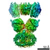
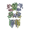
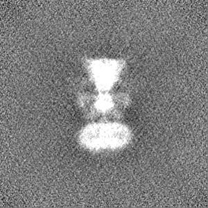
Journal: Sci Rep / Year: 2019 Title: Structural and Functional Insights into GluK3-kainate Receptor Desensitization and Recovery. Authors: Jyoti Kumari / Rajesh Vinnakota / Janesh Kumar / Abstract: GluK3-kainate receptors are atypical members of the iGluR family that reside at both the pre- and postsynapse and play a vital role in the regulation of synaptic transmission. For a better ...GluK3-kainate receptors are atypical members of the iGluR family that reside at both the pre- and postsynapse and play a vital role in the regulation of synaptic transmission. For a better understanding of structural changes that underlie receptor functions, GluK3 receptors were trapped in desensitized and resting/closed states and structures analyzed using single particle cryo-electron microscopy. While the desensitized GluK3 has domain organization as seen earlier for another kainate receptor-GluK2, antagonist bound GluK3 trapped a resting state with only two LBD domains in dimeric arrangement necessary for receptor activation. Using structures as a guide, we show that the N-linked glycans at the interface of GluK3 ATD and LBD likely mediate inter-domain interactions and attune receptor-gating properties. The mutational analysis also identified putative N-glycan interacting residues. Our results provide a molecular framework for understanding gating properties unique to GluK3 and exploring the role of N-linked glycosylation in their modulation.
History
Deposition
Feb 13, 2019
-
Header (metadata) release
Jul 24, 2019
-
Map release
Jul 24, 2019
-
Update
Nov 20, 2024
-
Current status
Nov 20, 2024
Processing site: PDBj / Status: Released
-
Structure visualization
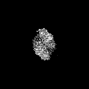
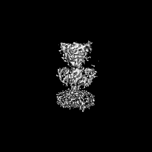
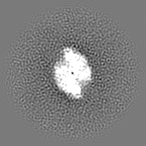
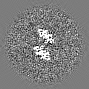
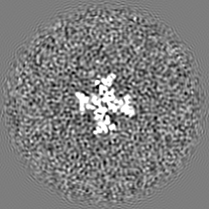
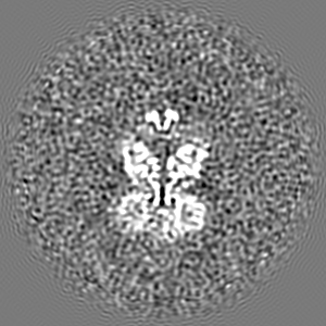
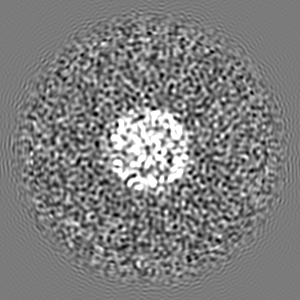
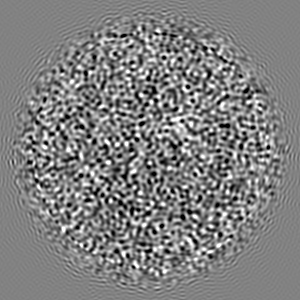
Movie
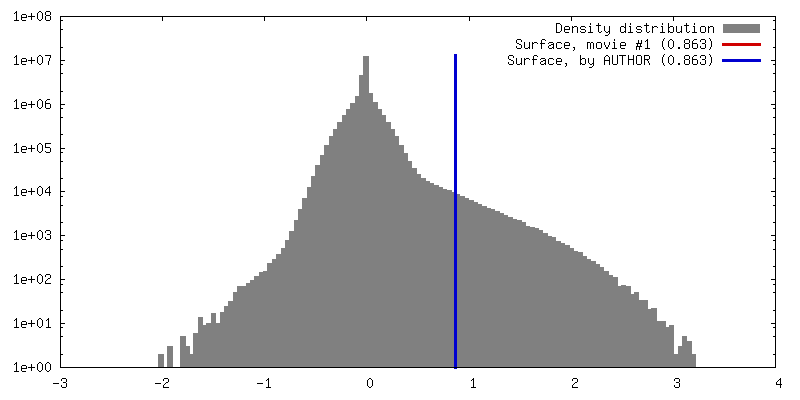
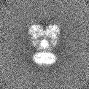
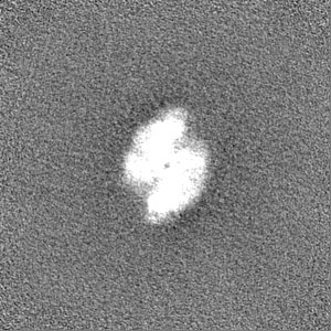
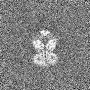
Surface view with section colored by density value
Purified and detergent solubilized GluK3 receptors
-
Electron microscopy
Microscope
FEI TITAN KRIOS
Image recording
Film or detector model: FEI FALCON III (4k x 4k) / Detector mode: COUNTING / Number grids imaged: 1 / Number real images: 719 / Average exposure time: 60.0 sec. / Average electron dose: 16.73 e/Å2
Electron beam
Acceleration voltage: 300 kV / Electron source: FIELD EMISSION GUN
Electron optics
Illumination mode: SPOT SCAN / Imaging mode: BRIGHT FIELD / Cs: 2.0 mm
Number classes used: 1 / Applied symmetry - Point group: C1 (asymmetric) / Algorithm: FOURIER SPACE / Resolution.type: BY AUTHOR / Resolution: 7.4 Å / Resolution method: FSC 0.143 CUT-OFF / Software - Name: cryoSPARC (ver. 1) / Software - details: final reconstruction / Number images used: 9730
Initial angle assignment
Type: COMMON LINE / Software - Name: cryoSPARC (ver. 1) / Software - details: Abinitio 3D reconstruction / Details: Abinitio 3D reconstruction module of cryosparc
In the structure databanks used in Yorodumi, some data are registered as the other names, "COVID-19 virus" and "2019-nCoV". Here are the details of the virus and the list of structure data.
Jan 31, 2019. EMDB accession codes are about to change! (news from PDBe EMDB page)
EMDB accession codes are about to change! (news from PDBe EMDB page)
The allocation of 4 digits for EMDB accession codes will soon come to an end. Whilst these codes will remain in use, new EMDB accession codes will include an additional digit and will expand incrementally as the available range of codes is exhausted. The current 4-digit format prefixed with “EMD-” (i.e. EMD-XXXX) will advance to a 5-digit format (i.e. EMD-XXXXX), and so on. It is currently estimated that the 4-digit codes will be depleted around Spring 2019, at which point the 5-digit format will come into force.
The EM Navigator/Yorodumi systems omit the EMD- prefix.
Related info.:Q: What is EMD? / ID/Accession-code notation in Yorodumi/EM Navigator
Yorodumi is a browser for structure data from EMDB, PDB, SASBDB, etc.
This page is also the successor to EM Navigator detail page, and also detail information page/front-end page for Omokage search.
The word "yorodu" (or yorozu) is an old Japanese word meaning "ten thousand". "mi" (miru) is to see.
Related info.:EMDB / PDB / SASBDB / Comparison of 3 databanks / Yorodumi Search / Aug 31, 2016. New EM Navigator & Yorodumi / Yorodumi Papers / Jmol/JSmol / Function and homology information / Changes in new EM Navigator and Yorodumi
 Movie
Movie Controller


 Open data
 Basic information
 Map data
 Sample
 Keywords
 Function and homology information
 Authors
 India, 2 items
  Citation
 Structure visualization
 Downloads & links
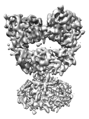 emd_9821.png
 http://ftp.pdbj.org/pub/emdb/structures/EMD-9821


 Z (Sec.)
 Y (Row.)
 X (Col.)


 Sample components
 Homo sapiens (human)
 Processing
 Electron microscopy
 FIELD EMISSION GUN

