 Movie
Movie Controller

[English] 日本語
 Yorodumi
- PDB-1qzw: Crystal structure of the complete core of archaeal SRP and implic... -
+ Open data

- Basic information
| Entry | Database: PDB / ID: 1qzw | ||||||
|---|---|---|---|---|---|---|---|
| Title | Crystal structure of the complete core of archaeal SRP and implications for inter-domain communication | ||||||
 Components Components |
| ||||||
 Keywords Keywords | Signaling Protein/RNA / Signal recognition particle / SRP / ribonucleoprotein complex / protein-RNA complex / protein targeting / Signaling Protein-RNA COMPLEX | ||||||
| Function / homology |  Function and homology information Function and homology informationsignal recognition particle / signal-recognition-particle GTPase / 7S RNA binding / SRP-dependent cotranslational protein targeting to membrane / GTPase activity / GTP binding Similarity search - Function | ||||||
| Biological species |   Sulfolobus solfataricus (archaea) Sulfolobus solfataricus (archaea) | ||||||
| Method |  X-RAY DIFFRACTION / SYNCHROTRON / MOLECULAR REPLACEMENT / Resolution: 4.1 Å X-RAY DIFFRACTION / SYNCHROTRON / MOLECULAR REPLACEMENT / Resolution: 4.1 Å | ||||||
 Authors Authors | Rosendal, K.R. / Wild, K. / Montoya, G. / Sinning, I. | ||||||
 Citation Citation | Journal: Proc.Natl.Acad.Sci.USA / Year: 2003 Title: Crystal structure of the complete core of archaeal signal recognition particle and implications for interdomain communication Authors: Rosendal, K.R. / Wild, K. / Montoya, G. / Sinning, I. | ||||||
| History |
| ||||||
| Remark 650 | HELIX DETERMINED METHOD: Authors determined | ||||||
| Remark 700 | SHEET DETERMINED METHOD: Authors determined |
- Structure visualization
| Structure viewer | Molecule: MolmilJmol/JSmol |
|---|
- Downloads & links
-Download
| PDBx/mmCIF format | 1qzw.cif.gz | 388.9 KB | Display | PDBx/mmCIF format |
|---|---|---|---|---|
| PDB format | pdb1qzw.ent.gz | 301.3 KB | Display | PDB format |
| PDBx/mmJSON format | 1qzw.json.gz | Tree view | PDBx/mmJSON format | |
| Others |  Other downloads Other downloads |
-Validation report
| Arichive directory | https://data.pdbj.org/pub/pdb/validation_reports/qz/1qzwftp://data.pdbj.org/pub/pdb/validation_reports/qz/1qzw | HTTPS FTP |
|---|
-Related structure data
| Related structure data |  1qzxC  1j8mS S: Starting model for refinement C: citing same article ( |
|---|---|
| Similar structure data |


-Links
 PDBj
PDBj


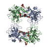
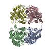
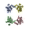
- Assembly
| Deposited unit | 
| ||||||||||||||||||||
|---|---|---|---|---|---|---|---|---|---|---|---|---|---|---|---|---|---|---|---|---|---|
| 1 | 
| ||||||||||||||||||||
| 2 | 
| ||||||||||||||||||||
| 3 | 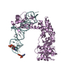
| ||||||||||||||||||||
| 4 | 
| ||||||||||||||||||||
| Unit cell |
| ||||||||||||||||||||
| Noncrystallographic symmetry (NCS) | NCS oper:
|
-Components
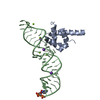
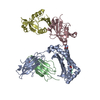
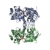
| #1: RNA chain | Mass: 15555.170 Da / Num. of mol.: 4 / Fragment: SRP RNA helix 8 Source method: isolated from a genetically manipulated source Source: (gene. exp.) Sulfolobus solfataricus (archaea) / Description: in vitro translation / Gene: 7S RNA / Plasmid: pUC18#2: Protein | Mass: 49463.430 Da / Num. of mol.: 4 Source method: isolated from a genetically manipulated source Source: (gene. exp.) Sulfolobus solfataricus (archaea) / Gene: SRP54 / Plasmid: pET24d / Production host:  |
|---|
-Experimental details
-Experiment
| Experiment | Method: X-RAY DIFFRACTION / Number of used crystals: 1 |
|---|
- Sample preparation
| Crystal | Density Matthews: 6.49 Å3/Da / Density % sol: 81.04 % | ||||||||||||||||||||||||||||||||||||||||||||||||||||||||
|---|---|---|---|---|---|---|---|---|---|---|---|---|---|---|---|---|---|---|---|---|---|---|---|---|---|---|---|---|---|---|---|---|---|---|---|---|---|---|---|---|---|---|---|---|---|---|---|---|---|---|---|---|---|---|---|---|---|
| Crystal grow | Temperature: 291 K / Method: vapor diffusion, hanging drop / pH: 4.5 Details: Ammonium sulphate, sodium chloride, sodium acetate, pH 4.5, VAPOR DIFFUSION, HANGING DROP, temperature 291K | ||||||||||||||||||||||||||||||||||||||||||||||||||||||||
| Components of the solutions |
| ||||||||||||||||||||||||||||||||||||||||||||||||||||||||
| Crystal grow | *PLUS Method: vapor diffusion, hanging dropDetails: Rosendal, K.R., (2004) Acta Crystallogr.,Sect.D, 60, 140. | ||||||||||||||||||||||||||||||||||||||||||||||||||||||||
| Components of the solutions | *PLUS
|
-Data collection
| Diffraction | Mean temperature: 100 K |
|---|---|
| Diffraction source | Source: SYNCHROTRON / Site: ESRF  / Beamline: ID29 / Wavelength: 0.9763 Å / Beamline: ID29 / Wavelength: 0.9763 Å |
| Detector | Type: ADSC QUANTUM 4 / Detector: CCD / Date: Apr 14, 2003 |
| Radiation | Protocol: SINGLE WAVELENGTH / Monochromatic (M) / Laue (L): M / Scattering type: x-ray |
| Radiation wavelength | Wavelength: 0.9763 Å / Relative weight: 1 |
| Reflection | Resolution: 4.1→30 Å / Num. obs: 50447 / % possible obs: 98.1 % / Observed criterion σ(F): 0 / Observed criterion σ(I): 0 / Redundancy: 1.9 % / Rsym value: 0.087 / Net I/σ(I): 4 |
| Reflection shell | Resolution: 4.1→4.32 Å / Redundancy: 1.9 % / Mean I/σ(I) obs: 1.5 / Num. unique all: 7462 / Rsym value: 0.481 / % possible all: 99.4 |
| Reflection | *PLUS Num. obs: 49660 / % possible obs: 97.2 % / Rmerge(I) obs: 0.087 |
| Reflection shell | *PLUS Lowest resolution: 4.31 Å / % possible obs: 98.1 % / Rmerge(I) obs: 0.481 |
- Processing
| Software |
| ||||||||||||||||||||||||||||||||||||||||||||||||||||||
|---|---|---|---|---|---|---|---|---|---|---|---|---|---|---|---|---|---|---|---|---|---|---|---|---|---|---|---|---|---|---|---|---|---|---|---|---|---|---|---|---|---|---|---|---|---|---|---|---|---|---|---|---|---|---|---|
| Refinement | Method to determine structure: MOLECULAR REPLACEMENT Starting model: PDB ID 1J8M Resolution: 4.1→30 Å / Rfactor Rfree error: 0.005 / Data cutoff high absF: 1576917.54 / Data cutoff high rms absF: 1576917.54 / Data cutoff low absF: 0 / Isotropic thermal model: OVERALL / Cross valid method: THROUGHOUT / σ(F): 0 / σ(I): 0 / Stereochemistry target values: Engh & Huber Details: The structure factor data associated with this file is from twinned crystal. To use structure factor data, this data will have to be detwinned.
| ||||||||||||||||||||||||||||||||||||||||||||||||||||||
| Solvent computation | Solvent model: FLAT MODEL / Bsol: 52.4117 Å2 / ksol: 0.105512 e/Å3 | ||||||||||||||||||||||||||||||||||||||||||||||||||||||
| Displacement parameters | Biso mean: 69.4 Å2
| ||||||||||||||||||||||||||||||||||||||||||||||||||||||
| Refine analyze |
| ||||||||||||||||||||||||||||||||||||||||||||||||||||||
| Refinement step | Cycle: LAST / Resolution: 4.1→30 Å
| ||||||||||||||||||||||||||||||||||||||||||||||||||||||
| Refine LS restraints |
| ||||||||||||||||||||||||||||||||||||||||||||||||||||||
| LS refinement shell | Refine-ID: X-RAY DIFFRACTION / Total num. of bins used: 6
| ||||||||||||||||||||||||||||||||||||||||||||||||||||||
| Xplor file |
| ||||||||||||||||||||||||||||||||||||||||||||||||||||||
| Refinement | *PLUS Highest resolution: 4.1 Å / Lowest resolution: 30 Å / % reflection Rfree: 9 % / Rfactor Rfree: 0.387 / Rfactor Rwork: 0.34 | ||||||||||||||||||||||||||||||||||||||||||||||||||||||
| Solvent computation | *PLUS | ||||||||||||||||||||||||||||||||||||||||||||||||||||||
| Displacement parameters | *PLUS | ||||||||||||||||||||||||||||||||||||||||||||||||||||||
| Refine LS restraints | *PLUS
|
