 Movie
Movie Controller

[English] 日本語
 Yorodumi
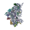
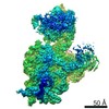
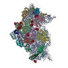
- EMDB-2685: Density map of GluK2 desensitized state in complex with 2S,4R-4-m... -
+ Open data

- Basic information
| Entry | Database: EMDB / ID: EMD-2685 | |||||||||
|---|---|---|---|---|---|---|---|---|---|---|
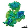
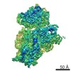
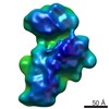
| Title | Density map of GluK2 desensitized state in complex with 2S,4R-4-methylglutamate | |||||||||
 Map data Map data | Reconstruction of GluK2 desensitized state | |||||||||
 Sample Sample |
| |||||||||
 Keywords Keywords | Membrane Protein / Ion Channel / Glutamate Receptor | |||||||||
| Function / homology |  Function and homology information Function and homology informationmossy fiber rosette / detection of cold stimulus involved in thermoception / Activation of Na-permeable kainate receptors / regulation of short-term neuronal synaptic plasticity / Activation of Ca-permeable Kainate Receptor / kainate selective glutamate receptor complex / ubiquitin conjugating enzyme binding / glutamate receptor activity / negative regulation of synaptic transmission, glutamatergic / regulation of JNK cascade ...mossy fiber rosette / detection of cold stimulus involved in thermoception / Activation of Na-permeable kainate receptors / regulation of short-term neuronal synaptic plasticity / Activation of Ca-permeable Kainate Receptor / kainate selective glutamate receptor complex / ubiquitin conjugating enzyme binding / glutamate receptor activity / negative regulation of synaptic transmission, glutamatergic / regulation of JNK cascade / inhibitory postsynaptic potential / receptor clustering / kainate selective glutamate receptor activity / extracellularly glutamate-gated ion channel activity / modulation of excitatory postsynaptic potential / ionotropic glutamate receptor complex / behavioral fear response / positive regulation of synaptic transmission / neuronal action potential / glutamate-gated receptor activity / glutamate-gated calcium ion channel activity / dendrite cytoplasm / ligand-gated monoatomic ion channel activity involved in regulation of presynaptic membrane potential / presynaptic modulation of chemical synaptic transmission / excitatory postsynaptic potential / hippocampal mossy fiber to CA3 synapse / SNARE binding / PDZ domain binding / regulation of long-term neuronal synaptic plasticity / synaptic transmission, glutamatergic / regulation of membrane potential / transmitter-gated monoatomic ion channel activity involved in regulation of postsynaptic membrane potential / intracellular protein transport / postsynaptic density membrane / modulation of chemical synaptic transmission / intracellular calcium ion homeostasis / terminal bouton / positive regulation of neuron apoptotic process / neuron apoptotic process / presynaptic membrane / scaffold protein binding / chemical synaptic transmission / negative regulation of neuron apoptotic process / perikaryon / postsynaptic membrane / postsynaptic density / axon / neuronal cell body / ubiquitin protein ligase binding / dendrite / synapse / glutamatergic synapse / membrane / identical protein binding / plasma membrane Similarity search - Function | |||||||||
| Biological species |  | |||||||||
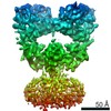
| Method | single particle reconstruction / cryo EM / Resolution: 7.6 Å | |||||||||
 Authors Authors | Meyerson JR / Kumar J / Chittori S / Rao P / Pierson J / Bartesaghi A / Mayer ML / Subramaniam S | |||||||||
 Citation Citation | Journal: Nature / Year: 2014 Title: Structural mechanism of glutamate receptor activation and desensitization. Authors: Joel R Meyerson / Janesh Kumar / Sagar Chittori / Prashant Rao / Jason Pierson / Alberto Bartesaghi / Mark L Mayer / Sriram Subramaniam /  Abstract: Ionotropic glutamate receptors are ligand-gated ion channels that mediate excitatory synaptic transmission in the vertebrate brain. To gain a better understanding of how structural changes gate ion ...Ionotropic glutamate receptors are ligand-gated ion channels that mediate excitatory synaptic transmission in the vertebrate brain. To gain a better understanding of how structural changes gate ion flux across the membrane, we trapped rat AMPA (α-amino-3-hydroxy-5-methyl-4-isoxazole propionic acid) and kainate receptor subtypes in their major functional states and analysed the resulting structures using cryo-electron microscopy. We show that transition to the active state involves a 'corkscrew' motion of the receptor assembly, driven by closure of the ligand-binding domain. Desensitization is accompanied by disruption of the amino-terminal domain tetramer in AMPA, but not kainate, receptors with a two-fold to four-fold symmetry transition in the ligand-binding domains in both subtypes. The 7.6 Å structure of a desensitized kainate receptor shows how these changes accommodate channel closing. These findings integrate previous physiological, biochemical and structural analyses of glutamate receptors and provide a molecular explanation for key steps in receptor gating. | |||||||||
| History |
|
- Structure visualization
| Movie |
Movie viewer |
|---|---|
| Structure viewer | EM map: SurfViewMolmilJmol/JSmol |
| Supplemental images |

- Downloads & links
-EMDB archive
| Map data | emd_2685.map.gz | 3.6 MB | EMDB map data format | |
|---|---|---|---|---|
| Header (meta data) | emd-2685-v30.xmlemd-2685.xml | 9.7 KB 9.7 KB | Display Display | EMDB header |
| Images | 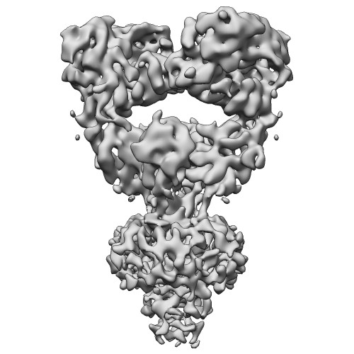 EMD-2685-GluK2-SYM.jpg EMD-2685-GluK2-SYM.jpg | 48.5 KB | ||
| Archive directory |  http://ftp.pdbj.org/pub/emdb/structures/EMD-2685ftp://ftp.pdbj.org/pub/emdb/structures/EMD-2685 http://ftp.pdbj.org/pub/emdb/structures/EMD-2685ftp://ftp.pdbj.org/pub/emdb/structures/EMD-2685 | HTTPS FTP |
-Related structure data
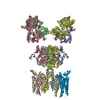
| Related structure data | 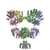 4uqqMC 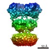 2680C 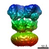 2684C 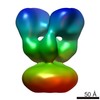 2686C 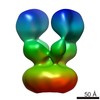 2687C 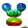 2688C 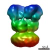 2689C 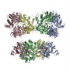 4uq6C 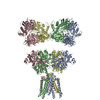 4uqjC 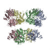 4uqkC M: atomic model generated by this map C: citing same article ( |
|---|---|
| Similar structure data |
-Links
| EMDB pages | EMDB (EBI/PDBe) / EMDataResource |
|---|---|
| Related items in Molecule of the Month |


-Map
| File | Download / File: emd_2685.map.gz / Format: CCP4 / Size: 29.8 MB / Type: IMAGE STORED AS FLOATING POINT NUMBER (4 BYTES) | ||||||||||||||||||||||||||||||||||||||||||||||||||||||||||||||||||||
|---|---|---|---|---|---|---|---|---|---|---|---|---|---|---|---|---|---|---|---|---|---|---|---|---|---|---|---|---|---|---|---|---|---|---|---|---|---|---|---|---|---|---|---|---|---|---|---|---|---|---|---|---|---|---|---|---|---|---|---|---|---|---|---|---|---|---|---|---|---|
| Annotation | Reconstruction of GluK2 desensitized state | ||||||||||||||||||||||||||||||||||||||||||||||||||||||||||||||||||||
| Projections & slices | Image control
Images are generated by Spider. | ||||||||||||||||||||||||||||||||||||||||||||||||||||||||||||||||||||
| Voxel size | X=Y=Z: 1.406 Å | ||||||||||||||||||||||||||||||||||||||||||||||||||||||||||||||||||||
| Density |
| ||||||||||||||||||||||||||||||||||||||||||||||||||||||||||||||||||||
| Symmetry | Space group: 1 | ||||||||||||||||||||||||||||||||||||||||||||||||||||||||||||||||||||
| Details | EMDB XML:
CCP4 map header:
| ||||||||||||||||||||||||||||||||||||||||||||||||||||||||||||||||||||
 Z (Sec.)
 Y (Row.)
 X (Col.)


-Supplemental data
- Sample components
-Entire : GluAK2 with 2S,4R-4-methylglutamate
| Entire | Name: GluAK2 with 2S,4R-4-methylglutamate |
|---|---|
| Components |
|
-Supramolecule #1000: GluAK2 with 2S,4R-4-methylglutamate
| Supramolecule | Name: GluAK2 with 2S,4R-4-methylglutamate / type: sample / ID: 1000 / Number unique components: 2 |
|---|---|
| Molecular weight | Experimental: 400 KDa / Theoretical: 400 KDa |
-Macromolecule #1: GluK2
| Macromolecule | Name: GluK2 / type: protein_or_peptide / ID: 1 / Oligomeric state: Tetramer / Recombinant expression: Yes |
|---|---|
| Source (natural) | Organism: |
| Molecular weight | Experimental: 400 KDa / Theoretical: 400 KDa |
| Recombinant expression | Organism:   Spodoptera frugiperda (fall armyworm) / Recombinant cell: Sf9 / Recombinant plasmid: pFastBac1 Spodoptera frugiperda (fall armyworm) / Recombinant cell: Sf9 / Recombinant plasmid: pFastBac1 |
| Sequence | UniProtKB: Glutamate receptor ionotropic, kainate 2 |
-Macromolecule #2: 2S,4R-4-methylglutamate
| Macromolecule | Name: 2S,4R-4-methylglutamate / type: ligand / ID: 2 / Recombinant expression: No |
|---|---|
| Source (natural) | Organism: synthetic construct (others) |
| Chemical component information | 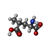 ChemComp-SYM: |
-Experimental details
-Structure determination
| Method | cryo EM |
|---|---|
 Processing Processing | single particle reconstruction |
| Aggregation state | particle |
-Sample preparation
| Concentration | 1.8 mg/mL |
|---|---|
| Buffer | pH: 8 Details: 150 mM NaCl, 20 mM Tris pH 8.0, 0.75 mM DDM, 1 mM 2S,4R-4-methylglutamate |
| Grid | Details: Vitrified specimens were prepared by adding 2.5 uL of liganded protein at 1.8 mg/ml to R2/2 holey carbon grids (Quantifoil, Jena, Germany) rendered hydrophilic by chemical treatment to ...Details: Vitrified specimens were prepared by adding 2.5 uL of liganded protein at 1.8 mg/ml to R2/2 holey carbon grids (Quantifoil, Jena, Germany) rendered hydrophilic by chemical treatment to enable particle distribution into the holes (Meyerson JR, Rao P, Kumar K, Chittori S, Banerjee S, Pierson J, Mayer ML, and Subramaniam S, manuscript in preparation). |
| Vitrification | Cryogen name: ETHANE / Chamber humidity: 100 % / Chamber temperature: 120 K / Instrument: FEI VITROBOT MARK IV / Method: Blot for 2 seconds before plunging |
- Electron microscopy
| Microscope | FEI TITAN KRIOS |
|---|---|
| Date | Aug 1, 2013 |
| Image recording | Category: CCD / Film or detector model: FEI FALCON II (4k x 4k) / Number real images: 4837 / Average electron dose: 25 e/Å2 |
| Electron beam | Acceleration voltage: 300 kV / Electron source:  FIELD EMISSION GUN FIELD EMISSION GUN |
| Electron optics | Calibrated magnification: 47000 / Illumination mode: FLOOD BEAM / Imaging mode: BRIGHT FIELD / Nominal defocus max: 3.5 µm / Nominal defocus min: 2.0 µm / Nominal magnification: 47000 |
| Sample stage | Specimen holder model: FEI TITAN KRIOS AUTOGRID HOLDER |
| Experimental equipment |  Model: Titan Krios / Image courtesy: FEI Company |
-Image processing
| Details | Particles were selected interactively at the computer terminal |
|---|---|
| CTF correction | Details: Each particle |
| Final reconstruction | Applied symmetry - Point group: C2 (2 fold cyclic) / Resolution.type: BY AUTHOR / Resolution: 7.6 Å / Resolution method: OTHER / Software - Name: Relion / Number images used: 21360 |
