 Movie
Movie Controller

[English] 日本語
 Yorodumi
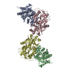
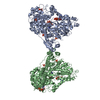
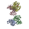
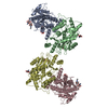
- PDB-3olz: Crystal structure of the GluK3 (GluR7) ATD dimer at 2.75 Angstrom... -
+ Open data

- Basic information
| Entry | Database: PDB / ID: 3olz | ||||||
|---|---|---|---|---|---|---|---|
| Title | Crystal structure of the GluK3 (GluR7) ATD dimer at 2.75 Angstrom resolution | ||||||
 Components Components | Glutamate receptor, ionotropic kainate 3 | ||||||
 Keywords Keywords | MEMBRANE PROTEIN / Ion channel | ||||||
| Function / homology |  Function and homology information Function and homology informationPresynaptic function of Kainate receptors / cochlear hair cell ribbon synapse / adenylate cyclase inhibiting G protein-coupled glutamate receptor activity / G protein-coupled glutamate receptor signaling pathway / Activation of Ca-permeable Kainate Receptor / kainate selective glutamate receptor complex / glutamate receptor activity / negative regulation of synaptic transmission, glutamatergic / glutamate receptor signaling pathway / kainate selective glutamate receptor activity ...Presynaptic function of Kainate receptors / cochlear hair cell ribbon synapse / adenylate cyclase inhibiting G protein-coupled glutamate receptor activity / G protein-coupled glutamate receptor signaling pathway / Activation of Ca-permeable Kainate Receptor / kainate selective glutamate receptor complex / glutamate receptor activity / negative regulation of synaptic transmission, glutamatergic / glutamate receptor signaling pathway / kainate selective glutamate receptor activity / glutamate-gated receptor activity / glutamate-gated calcium ion channel activity / dendrite cytoplasm / ligand-gated monoatomic ion channel activity involved in regulation of presynaptic membrane potential / synaptic transmission, glutamatergic / regulation of membrane potential / transmitter-gated monoatomic ion channel activity involved in regulation of postsynaptic membrane potential / postsynaptic density membrane / modulation of chemical synaptic transmission / terminal bouton / presynaptic membrane / chemical synaptic transmission / perikaryon / postsynaptic membrane / axon / dendrite / glutamatergic synapse / plasma membrane Similarity search - Function | ||||||
| Biological species |  | ||||||
| Method |  X-RAY DIFFRACTION / SYNCHROTRON / MOLECULAR REPLACEMENT / Resolution: 2.75 Å X-RAY DIFFRACTION / SYNCHROTRON / MOLECULAR REPLACEMENT / Resolution: 2.75 Å | ||||||
 Authors Authors | Kumar, J. / Mayer, M.L. | ||||||
 Citation Citation | Journal: J.Mol.Biol. / Year: 2010 Title: Crystal Structures of the Glutamate Receptor Ion Channel GluK3 and GluK5 Amino-Terminal Domains. Authors: Kumar, J. / Mayer, M.L. | ||||||
| History |
|
- Structure visualization
| Structure viewer | Molecule: MolmilJmol/JSmol |
|---|
- Downloads & links
-Download
| PDBx/mmCIF format | 3olz.cif.gz | 443.2 KB | Display | PDBx/mmCIF format |
|---|---|---|---|---|
| PDB format | pdb3olz.ent.gz | 374.4 KB | Display | PDB format |
| PDBx/mmJSON format | 3olz.json.gz | Tree view | PDBx/mmJSON format | |
| Others |  Other downloads Other downloads |
-Validation report
| Arichive directory | https://data.pdbj.org/pub/pdb/validation_reports/ol/3olzftp://data.pdbj.org/pub/pdb/validation_reports/ol/3olz | HTTPS FTP |
|---|
-Related structure data
| Related structure data |  3om0C  3om1C  3h6gS S: Starting model for refinement C: citing same article ( |
|---|---|
| Similar structure data |


-Links
 PDBj
PDBj

- Assembly
| Deposited unit | 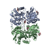
| ||||||||
|---|---|---|---|---|---|---|---|---|---|
| 1 | 
| ||||||||
| 2 |
| ||||||||
| Unit cell |
|
-Components
| #1: Protein | Mass: 45352.035 Da / Num. of mol.: 2 Source method: isolated from a genetically manipulated source Source: (gene. exp.)  Homo Sapiens (human) / References: UniProt: D3ZDH2, UniProt: P42264*PLUS Homo Sapiens (human) / References: UniProt: D3ZDH2, UniProt: P42264*PLUS#2: Sugar | ChemComp-NAG /   Type: D-saccharide, beta linking / Mass: 221.208 Da / Num. of mol.: 5 Type: D-saccharide, beta linking / Mass: 221.208 Da / Num. of mol.: 5Source method: isolated from a genetically manipulated source Formula: C8H15NO6 #3: Chemical |   Mass: 35.453 Da / Num. of mol.: 2 / Source method: obtained synthetically / Formula: Cl Mass: 35.453 Da / Num. of mol.: 2 / Source method: obtained synthetically / Formula: Cl#4: Water | ChemComp-HOH / |  Mass: 18.015 Da / Num. of mol.: 14 / Source method: isolated from a natural source / Formula: H2O Mass: 18.015 Da / Num. of mol.: 14 / Source method: isolated from a natural source / Formula: H2OHas protein modification | Y | Sequence details | THE SEQUENCE CORRESPONDS TO GLUTAMATE RECEPTOR SUBUNIT KAINATE SUBTYPE [RATTUS NORVEGICUS], GENE ...THE SEQUENCE CORRESPOND | |
|---|
-Experimental details
-Experiment
| Experiment | Method: X-RAY DIFFRACTION / Number of used crystals: 1 |
|---|
- Sample preparation
| Crystal | Density Matthews: 3.18 Å3/Da / Density % sol: 61.36 % |
|---|---|
| Crystal grow | Temperature: 277 K / Method: vapor diffusion, hanging drop / pH: 6.8 Details: 0.1 M MgCl2, 0.1 M MES, 10% Isopropanol, 5% PEG 4K, pH 6.8, VAPOR DIFFUSION, HANGING DROP, temperature 277K |
-Data collection
| Diffraction | Mean temperature: 100 K |
|---|---|
| Diffraction source | Source: SYNCHROTRON / Site: APS  / Beamline: 23-ID-B / Wavelength: 1.0332 Å / Beamline: 23-ID-B / Wavelength: 1.0332 Å |
| Detector | Type: MARMOSAIC 300 mm CCD / Detector: CCD / Date: Nov 24, 2009 |
| Radiation | Monochromator: Si(111) Double Crystal Monochrometer / Protocol: SINGLE WAVELENGTH / Monochromatic (M) / Laue (L): M / Scattering type: x-ray |
| Radiation wavelength | Wavelength: 1.0332 Å / Relative weight: 1 |
| Reflection | Resolution: 2.75→45 Å / Num. all: 29967 / Num. obs: 29967 / % possible obs: 98 % / Observed criterion σ(F): 1 / Observed criterion σ(I): 1 / Redundancy: 4.5 % / Biso Wilson estimate: 54.8 Å2 / Rmerge(I) obs: 0.091 / Net I/σ(I): 16 |
| Reflection shell | Resolution: 2.75→2.85 Å / Redundancy: 4.4 % / Rmerge(I) obs: 0.64 / Mean I/σ(I) obs: 2.25 / Num. unique all: 2894 / % possible all: 97.3 |
- Processing
| Software |
| |||||||||||||||||||||||||||||||||||||||||||||||||||||||||||||||||||||||||||||||||||||||||||||||||||||||||||||||||||||||||||||||||||||||||||||||||||||||||||||||||||||||||||||||||||||||||||||||||||||||||||||||||||||||||||||||||
|---|---|---|---|---|---|---|---|---|---|---|---|---|---|---|---|---|---|---|---|---|---|---|---|---|---|---|---|---|---|---|---|---|---|---|---|---|---|---|---|---|---|---|---|---|---|---|---|---|---|---|---|---|---|---|---|---|---|---|---|---|---|---|---|---|---|---|---|---|---|---|---|---|---|---|---|---|---|---|---|---|---|---|---|---|---|---|---|---|---|---|---|---|---|---|---|---|---|---|---|---|---|---|---|---|---|---|---|---|---|---|---|---|---|---|---|---|---|---|---|---|---|---|---|---|---|---|---|---|---|---|---|---|---|---|---|---|---|---|---|---|---|---|---|---|---|---|---|---|---|---|---|---|---|---|---|---|---|---|---|---|---|---|---|---|---|---|---|---|---|---|---|---|---|---|---|---|---|---|---|---|---|---|---|---|---|---|---|---|---|---|---|---|---|---|---|---|---|---|---|---|---|---|---|---|---|---|---|---|---|---|---|---|---|---|---|---|---|---|---|---|---|---|---|---|---|---|
| Refinement | Method to determine structure: MOLECULAR REPLACEMENT Starting model: PDB ENTRY 3H6G Resolution: 2.75→43.307 Å / SU ML: 0.35 / Cross valid method: THROUGHOUT / σ(F): 1.34 / Stereochemistry target values: ML
| |||||||||||||||||||||||||||||||||||||||||||||||||||||||||||||||||||||||||||||||||||||||||||||||||||||||||||||||||||||||||||||||||||||||||||||||||||||||||||||||||||||||||||||||||||||||||||||||||||||||||||||||||||||||||||||||||
| Solvent computation | Shrinkage radii: 0.77 Å / VDW probe radii: 0.9 Å / Solvent model: FLAT BULK SOLVENT MODEL / Bsol: 49.177 Å2 / ksol: 0.406 e/Å3 | |||||||||||||||||||||||||||||||||||||||||||||||||||||||||||||||||||||||||||||||||||||||||||||||||||||||||||||||||||||||||||||||||||||||||||||||||||||||||||||||||||||||||||||||||||||||||||||||||||||||||||||||||||||||||||||||||
| Displacement parameters |
| |||||||||||||||||||||||||||||||||||||||||||||||||||||||||||||||||||||||||||||||||||||||||||||||||||||||||||||||||||||||||||||||||||||||||||||||||||||||||||||||||||||||||||||||||||||||||||||||||||||||||||||||||||||||||||||||||
| Refinement step | Cycle: LAST / Resolution: 2.75→43.307 Å
| |||||||||||||||||||||||||||||||||||||||||||||||||||||||||||||||||||||||||||||||||||||||||||||||||||||||||||||||||||||||||||||||||||||||||||||||||||||||||||||||||||||||||||||||||||||||||||||||||||||||||||||||||||||||||||||||||
| Refine LS restraints |
| |||||||||||||||||||||||||||||||||||||||||||||||||||||||||||||||||||||||||||||||||||||||||||||||||||||||||||||||||||||||||||||||||||||||||||||||||||||||||||||||||||||||||||||||||||||||||||||||||||||||||||||||||||||||||||||||||
| LS refinement shell |
| |||||||||||||||||||||||||||||||||||||||||||||||||||||||||||||||||||||||||||||||||||||||||||||||||||||||||||||||||||||||||||||||||||||||||||||||||||||||||||||||||||||||||||||||||||||||||||||||||||||||||||||||||||||||||||||||||
| Refinement TLS params. | Method: refined / Refine-ID: X-RAY DIFFRACTION
| |||||||||||||||||||||||||||||||||||||||||||||||||||||||||||||||||||||||||||||||||||||||||||||||||||||||||||||||||||||||||||||||||||||||||||||||||||||||||||||||||||||||||||||||||||||||||||||||||||||||||||||||||||||||||||||||||
| Refinement TLS group |
|