 Movie
Movie Controller

[English] 日本語
 Yorodumi
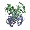
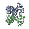
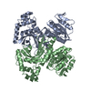
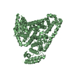
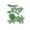
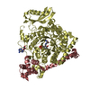
- PDB-1ebu: HOMOSERINE DEHYDROGENASE COMPLEX WITH NAD ANALOGUE AND L-HOMOSERINE -
+ Open data

- Basic information
| Entry | Database: PDB / ID: 1ebu | |||||||||
|---|---|---|---|---|---|---|---|---|---|---|
| Title | HOMOSERINE DEHYDROGENASE COMPLEX WITH NAD ANALOGUE AND L-HOMOSERINE | |||||||||
 Components Components | HOMOSERINE DEHYDROGENASE | |||||||||
 Keywords Keywords | OXIDOREDUCTASE / homoserine / dehydrogenase / dinucleotide / ternary / analogue | |||||||||
| Function / homology |  Function and homology information Function and homology informationaspartate family amino acid biosynthetic process / homoserine dehydrogenase / homoserine dehydrogenase activity / L-homoserine biosynthetic process / L-threonine biosynthetic process / : / NAD+ binding / NADP binding / nucleus / cytoplasm Similarity search - Function | |||||||||
| Biological species |  | |||||||||
| Method |  X-RAY DIFFRACTION / Resolution: 2.6 Å X-RAY DIFFRACTION / Resolution: 2.6 Å | |||||||||
 Authors Authors | DeLaBarre, B. / Thompson, P.R. / Wright, G.D. / Berghuis, A.M. | |||||||||
 Citation Citation | Journal: Nat.Struct.Biol. / Year: 2000 Title: Crystal structures of homoserine dehydrogenase suggest a novel catalytic mechanism for oxidoreductases. Authors: DeLaBarre, B. / Thompson, P.R. / Wright, G.D. / Berghuis, A.M. #1: Journal: Acta Crystallogr.,Sect.D / Year: 1998Title: Crystallization and preliminary X-ray diffraction studies of homoserine dehydrogenase from Saccharomyces cerevisiae Authors: DeLaBarre, B. / Jacques, S.L. / Pratt, C.E. / Wright, G.D. / Berghuis, A.M. | |||||||||
| History |
|
- Structure visualization
| Structure viewer | Molecule: MolmilJmol/JSmol |
|---|
- Downloads & links
-Download
| PDBx/mmCIF format | 1ebu.cif.gz | 282.2 KB | Display | PDBx/mmCIF format |
|---|---|---|---|---|
| PDB format | pdb1ebu.ent.gz | 229.1 KB | Display | PDB format |
| PDBx/mmJSON format | 1ebu.json.gz | Tree view | PDBx/mmJSON format | |
| Others |  Other downloads Other downloads |
-Validation report
| Arichive directory | https://data.pdbj.org/pub/pdb/validation_reports/eb/1ebuftp://data.pdbj.org/pub/pdb/validation_reports/eb/1ebu | HTTPS FTP |
|---|
-Related structure data


-Links
 PDBj
PDBj- Assembly
| Deposited unit | 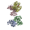
| ||||||||
|---|---|---|---|---|---|---|---|---|---|
| 1 | 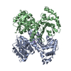
| ||||||||
| 2 | 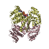
| ||||||||
| Unit cell |
| ||||||||
| Details | The asymmetric unit contains 2 dimers: A/B and C/D. The ternary complex is found in the D chain protomer |
-Components
| #1: Protein | Mass: 38413.766 Da / Num. of mol.: 4 Source method: isolated from a genetically manipulated source Source: (gene. exp.) Gene: HOM6P / Production host:  #2: Chemical | ChemComp-NA /   Mass: 22.990 Da / Num. of mol.: 4 / Source method: obtained synthetically / Formula: Na Mass: 22.990 Da / Num. of mol.: 4 / Source method: obtained synthetically / Formula: Na#3: Chemical | ChemComp-NDA / |   Mass: 649.442 Da / Num. of mol.: 1 / Source method: obtained synthetically / Formula: C21H29N7O13P2 Mass: 649.442 Da / Num. of mol.: 1 / Source method: obtained synthetically / Formula: C21H29N7O13P2#4: Chemical | ChemComp-HSE / | 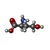  Type: L-peptide linking / Mass: 119.119 Da / Num. of mol.: 1 / Source method: obtained synthetically / Formula: C4H9NO3 Type: L-peptide linking / Mass: 119.119 Da / Num. of mol.: 1 / Source method: obtained synthetically / Formula: C4H9NO3#5: Water | ChemComp-HOH / |  Mass: 18.015 Da / Num. of mol.: 351 / Source method: isolated from a natural source / Formula: H2O Mass: 18.015 Da / Num. of mol.: 351 / Source method: isolated from a natural source / Formula: H2O |
|---|
-Experimental details
-Experiment
| Experiment | Method: X-RAY DIFFRACTION / Number of used crystals: 1 |
|---|
- Sample preparation
| Crystal | Density Matthews: 2.4 Å3/Da / Density % sol: 48.74 % | ||||||||||||||||||||||||||||||||||||||||||
|---|---|---|---|---|---|---|---|---|---|---|---|---|---|---|---|---|---|---|---|---|---|---|---|---|---|---|---|---|---|---|---|---|---|---|---|---|---|---|---|---|---|---|---|
| Crystal grow | Temperature: 295 K / Method: vapor diffusion, hanging drop / pH: 6.5 Details: PEG 8K, L-homoserine, 3-aminopyridine dinucleotide, sodium cacodylate , pH 6.5, VAPOR DIFFUSION, HANGING DROP, temperature 295K | ||||||||||||||||||||||||||||||||||||||||||
| Crystal grow | *PLUS pH: 8.5 | ||||||||||||||||||||||||||||||||||||||||||
| Components of the solutions | *PLUS
|
-Data collection
| Diffraction | Mean temperature: 218 K |
|---|---|
| Diffraction source | Source: ROTATING ANODE / Type: RIGAKU RU200 / Wavelength: 1.5418 |
| Detector | Type: RIGAKU RAXIS II / Detector: IMAGE PLATE / Date: Feb 22, 1996 |
| Radiation | Protocol: SINGLE WAVELENGTH / Monochromatic (M) / Laue (L): M / Scattering type: x-ray |
| Radiation wavelength | Wavelength: 1.5418 Å / Relative weight: 1 |
| Reflection | Resolution: 2.6→40 Å / Num. all: 44409 / Num. obs: 164716 / % possible obs: 99.2 % / Observed criterion σ(F): 3 / Observed criterion σ(I): 0 / Redundancy: 3.7 % / Biso Wilson estimate: 35.4 Å2 / Rmerge(I) obs: 0.081 / Net I/σ(I): 13.3 |
| Reflection shell | Resolution: 2.6→2.69 Å / Redundancy: 3.5 % / Rmerge(I) obs: 0.344 / Num. unique all: 4392 / % possible all: 98.1 |
| Reflection | *PLUS |
| Reflection shell | *PLUS % possible obs: 98.1 % |
- Processing
| Software |
| ||||||||||||||||||||||||||||||||||||||||||||||||||||||||||||||||||||||||||||||||
|---|---|---|---|---|---|---|---|---|---|---|---|---|---|---|---|---|---|---|---|---|---|---|---|---|---|---|---|---|---|---|---|---|---|---|---|---|---|---|---|---|---|---|---|---|---|---|---|---|---|---|---|---|---|---|---|---|---|---|---|---|---|---|---|---|---|---|---|---|---|---|---|---|---|---|---|---|---|---|---|---|---|
| Refinement | Resolution: 2.6→39.4 Å / Rfactor Rfree error: 0.004 / Data cutoff high absF: 1625863.12 / Data cutoff low absF: 0 / Isotropic thermal model: RESTRAINED / Cross valid method: THROUGHOUT / σ(F): 2 / σ(I): 0 / Stereochemistry target values: Engh & Huber / Details: Model refined against amplitudes
| ||||||||||||||||||||||||||||||||||||||||||||||||||||||||||||||||||||||||||||||||
| Solvent computation | Solvent model: FLAT MODEL / Bsol: 34 Å2 / ksol: 0.322 e/Å3 | ||||||||||||||||||||||||||||||||||||||||||||||||||||||||||||||||||||||||||||||||
| Displacement parameters | Biso mean: 28.6 Å2
| ||||||||||||||||||||||||||||||||||||||||||||||||||||||||||||||||||||||||||||||||
| Refine analyze |
| ||||||||||||||||||||||||||||||||||||||||||||||||||||||||||||||||||||||||||||||||
| Refinement step | Cycle: LAST / Resolution: 2.6→39.4 Å
| ||||||||||||||||||||||||||||||||||||||||||||||||||||||||||||||||||||||||||||||||
| Refine LS restraints |
| ||||||||||||||||||||||||||||||||||||||||||||||||||||||||||||||||||||||||||||||||
| LS refinement shell | Resolution: 2.6→2.76 Å / Rfactor Rfree error: 0.016 / Total num. of bins used: 6
| ||||||||||||||||||||||||||||||||||||||||||||||||||||||||||||||||||||||||||||||||
| Xplor file |
| ||||||||||||||||||||||||||||||||||||||||||||||||||||||||||||||||||||||||||||||||
| Software | *PLUS Name: CNS / Version: 0.9 / Classification: refinement | ||||||||||||||||||||||||||||||||||||||||||||||||||||||||||||||||||||||||||||||||
| Refine LS restraints | *PLUS
|
