 Movie
Movie Controller

[English] 日本語
 Yorodumi
- PDB-1q7g: Homoserine Dehydrogenase in complex with suicide inhibitor comple... -
+ Open data

- Basic information
| Entry | Database: PDB / ID: 1q7g | ||||||
|---|---|---|---|---|---|---|---|
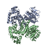
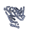
| Title | Homoserine Dehydrogenase in complex with suicide inhibitor complex NAD-5-hydroxy-4-Oxonorvaline | ||||||
 Components Components | Homoserine dehydrogenase | ||||||
 Keywords Keywords | OXIDOREDUCTASE | ||||||
| Function / homology |  Function and homology information Function and homology informationaspartate family amino acid biosynthetic process / homoserine dehydrogenase / homoserine dehydrogenase activity / L-homoserine biosynthetic process / L-threonine biosynthetic process / : / NAD+ binding / NADP binding / nucleus / cytoplasm Similarity search - Function | ||||||
| Biological species |  | ||||||
| Method |  X-RAY DIFFRACTION / SYNCHROTRON / MOLECULAR REPLACEMENT / Resolution: 2.6 Å X-RAY DIFFRACTION / SYNCHROTRON / MOLECULAR REPLACEMENT / Resolution: 2.6 Å | ||||||
 Authors Authors | Jacques, S.L. / Mirza, I.A. / Ejim, L. / Koteva, K. / Hughes, D.W. / Green, K. / Kinach, R. / Honek, J.F. / Lai, H.K. / Berghuis, A.M. / Wright, G.D. | ||||||
 Citation Citation | Journal: Chem.Biol. / Year: 2003 Title: Enzyme assisted suicide: Molecular basis for the antifungal activity of 5-hydroxy-4-oxonorvaline by potent inhibition of homoserine dehydrogenase Authors: Jacques, S.L. / Mirza, I.A. / Ejim, L. / Koteva, K. / Hughes, D.W. / Green, K. / Kinach, R. / Honek, J.F. / Lai, H.K. / Berghuis, A.M. / Wright, G.D. | ||||||
| History |
| ||||||
| Remark 600 | HETEROGEN Two possible adducts were constructed differing in chirality at the HON C5 position and ...HETEROGEN Two possible adducts were constructed differing in chirality at the HON C5 position and modeled into the active site. Subsequent electron density maps and monitoring of Rfree did not indicate a convincing preference for either configuration, and thus the current model describes the chirality of the HON C5 to be 50% S and 50% R. |
- Structure visualization
| Structure viewer | Molecule: MolmilJmol/JSmol |
|---|
- Downloads & links
-Download
| PDBx/mmCIF format | 1q7g.cif.gz | 147.5 KB | Display | PDBx/mmCIF format |
|---|---|---|---|---|
| PDB format | pdb1q7g.ent.gz | 116.3 KB | Display | PDB format |
| PDBx/mmJSON format | 1q7g.json.gz | Tree view | PDBx/mmJSON format | |
| Others |  Other downloads Other downloads |
-Validation report
| Arichive directory | https://data.pdbj.org/pub/pdb/validation_reports/q7/1q7gftp://data.pdbj.org/pub/pdb/validation_reports/q7/1q7g | HTTPS FTP |
|---|
-Related structure data
| Related structure data |  1ebfS S: Starting model for refinement |
|---|---|
| Similar structure data |


-Links
 PDBj
PDBj- Assembly
| Deposited unit | 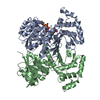
| ||||||||
|---|---|---|---|---|---|---|---|---|---|
| 1 |
| ||||||||
| Unit cell |
|
-Components
| #1: Protein | Mass: 38544.961 Da / Num. of mol.: 2 Source method: isolated from a genetically manipulated source Source: (gene. exp.) Gene: HOM6, YJR139C OR J2132 / Plasmid: pet22b / Production host:  #2: Chemical |   Mass: 22.990 Da / Num. of mol.: 2 / Source method: obtained synthetically / Formula: Na Mass: 22.990 Da / Num. of mol.: 2 / Source method: obtained synthetically / Formula: Na#3: Chemical | ChemComp-NHO / |   Mass: 809.546 Da / Num. of mol.: 1 / Source method: obtained synthetically / Formula: C26H35N8O18P2 Mass: 809.546 Da / Num. of mol.: 1 / Source method: obtained synthetically / Formula: C26H35N8O18P2#4: Water | ChemComp-HOH / |  Mass: 18.015 Da / Num. of mol.: 41 / Source method: isolated from a natural source / Formula: H2O Mass: 18.015 Da / Num. of mol.: 41 / Source method: isolated from a natural source / Formula: H2O |
|---|
-Experimental details
-Experiment
| Experiment | Method: X-RAY DIFFRACTION / Number of used crystals: 1 |
|---|
- Sample preparation
| Crystal | Density Matthews: 2.64 Å3/Da / Density % sol: 53.39 % | ||||||||||||||||||||||||||||||||||||||||||
|---|---|---|---|---|---|---|---|---|---|---|---|---|---|---|---|---|---|---|---|---|---|---|---|---|---|---|---|---|---|---|---|---|---|---|---|---|---|---|---|---|---|---|---|
| Crystal grow | Temperature: 295 K / Method: vapor diffusion, hanging drop / pH: 4.4 Details: Sodium Acetate, Ammonium Sulfate, pH 4.4, VAPOR DIFFUSION, HANGING DROP, temperature 295K | ||||||||||||||||||||||||||||||||||||||||||
| Crystal grow | *PLUS Method: vapor diffusion, hanging dropDetails: DeLaBarre, B., (1998) Acta Crystallogr., Sect.D, 54, 413. | ||||||||||||||||||||||||||||||||||||||||||
| Components of the solutions | *PLUS
|
-Data collection
| Diffraction | Mean temperature: 218 K |
|---|---|
| Diffraction source | Source: SYNCHROTRON / Site: NSLS  / Beamline: X8C / Wavelength: 1.0722 Å / Beamline: X8C / Wavelength: 1.0722 Å |
| Detector | Type: ADSC QUANTUM 4 / Detector: CCD / Date: Feb 28, 2001 |
| Radiation | Protocol: SINGLE WAVELENGTH / Monochromatic (M) / Laue (L): M / Scattering type: x-ray |
| Radiation wavelength | Wavelength: 1.0722 Å / Relative weight: 1 |
| Reflection | Resolution: 2.6→99 Å / Num. all: 24854 / Num. obs: 24854 / % possible obs: 94.6 % / Observed criterion σ(F): 0 / Observed criterion σ(I): 0 / Biso Wilson estimate: 20.7 Å2 / Rmerge(I) obs: 0.081 |
| Reflection shell | Resolution: 2.6→2.69 Å / Rmerge(I) obs: 0.257 / % possible all: 72 |
| Reflection shell | *PLUS % possible obs: 72 % / Mean I/σ(I) obs: 4 |
- Processing
| Software |
| ||||||||||||||||||||||||||||||||||||||||||||||||||||||||||||||||||||||||||||||||
|---|---|---|---|---|---|---|---|---|---|---|---|---|---|---|---|---|---|---|---|---|---|---|---|---|---|---|---|---|---|---|---|---|---|---|---|---|---|---|---|---|---|---|---|---|---|---|---|---|---|---|---|---|---|---|---|---|---|---|---|---|---|---|---|---|---|---|---|---|---|---|---|---|---|---|---|---|---|---|---|---|---|
| Refinement | Method to determine structure: MOLECULAR REPLACEMENT Starting model: PDB ENTRY 1EBF Resolution: 2.6→40.43 Å / Rfactor Rfree error: 0.006 / Data cutoff high absF: 125946.93 / Data cutoff high rms absF: 125946.93 / Data cutoff low absF: 0 / Isotropic thermal model: RESTRAINED / Cross valid method: THROUGHOUT / σ(F): 0 / Stereochemistry target values: Engh & Huber
| ||||||||||||||||||||||||||||||||||||||||||||||||||||||||||||||||||||||||||||||||
| Solvent computation | Solvent model: FLAT MODEL / Bsol: 37.9335 Å2 / ksol: 0.389643 e/Å3 | ||||||||||||||||||||||||||||||||||||||||||||||||||||||||||||||||||||||||||||||||
| Displacement parameters | Biso mean: 33.4 Å2
| ||||||||||||||||||||||||||||||||||||||||||||||||||||||||||||||||||||||||||||||||
| Refine analyze |
| ||||||||||||||||||||||||||||||||||||||||||||||||||||||||||||||||||||||||||||||||
| Refinement step | Cycle: LAST / Resolution: 2.6→40.43 Å
| ||||||||||||||||||||||||||||||||||||||||||||||||||||||||||||||||||||||||||||||||
| Refine LS restraints |
| ||||||||||||||||||||||||||||||||||||||||||||||||||||||||||||||||||||||||||||||||
| LS refinement shell | Resolution: 2.6→2.76 Å / Rfactor Rfree error: 0.022 / Total num. of bins used: 6
| ||||||||||||||||||||||||||||||||||||||||||||||||||||||||||||||||||||||||||||||||
| Xplor file |
| ||||||||||||||||||||||||||||||||||||||||||||||||||||||||||||||||||||||||||||||||
| Refinement | *PLUS Highest resolution: 2.6 Å / Rfactor Rfree: 0.295 / Rfactor Rwork: 0.232 | ||||||||||||||||||||||||||||||||||||||||||||||||||||||||||||||||||||||||||||||||
| Solvent computation | *PLUS | ||||||||||||||||||||||||||||||||||||||||||||||||||||||||||||||||||||||||||||||||
| Displacement parameters | *PLUS | ||||||||||||||||||||||||||||||||||||||||||||||||||||||||||||||||||||||||||||||||
| Refine LS restraints | *PLUS
|
