 Movie
Movie Controller

[English] 日本語
 Yorodumi
- PDB-3h6h: Crystal structure of the GluR6 amino terminal domain dimer assemb... -
+ Open data

- Basic information
| Entry | Database: PDB / ID: 3h6h | ||||||
|---|---|---|---|---|---|---|---|
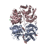
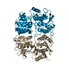
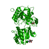
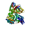
| Title | Crystal structure of the GluR6 amino terminal domain dimer assembly MPD form | ||||||
 Components Components | Glutamate receptor, ionotropic kainate 2 | ||||||
 Keywords Keywords | MEMBRANE PROTEIN / MEMBRANE PROTEIN GLYCOPROTEIN / Cell junction / Cell membrane / Glycoprotein / Ion transport / Ionic channel / Isopeptide bond / Membrane / Postsynaptic cell membrane / Receptor / RNA editing / Synapse / Transmembrane / Transport | ||||||
| Function / homology |  Function and homology information Function and homology informationmossy fiber rosette / detection of cold stimulus involved in thermoception / Activation of Na-permeable kainate receptors / Activation of Ca-permeable Kainate Receptor / kainate selective glutamate receptor complex / regulation of short-term neuronal synaptic plasticity / ubiquitin conjugating enzyme binding / glutamate receptor activity / negative regulation of synaptic transmission, glutamatergic / regulation of JNK cascade ...mossy fiber rosette / detection of cold stimulus involved in thermoception / Activation of Na-permeable kainate receptors / Activation of Ca-permeable Kainate Receptor / kainate selective glutamate receptor complex / regulation of short-term neuronal synaptic plasticity / ubiquitin conjugating enzyme binding / glutamate receptor activity / negative regulation of synaptic transmission, glutamatergic / regulation of JNK cascade / receptor clustering / inhibitory postsynaptic potential / kainate selective glutamate receptor activity / extracellularly glutamate-gated ion channel activity / modulation of excitatory postsynaptic potential / ionotropic glutamate receptor complex / positive regulation of synaptic transmission / behavioral fear response / neuronal action potential / glutamate-gated receptor activity / glutamate-gated calcium ion channel activity / dendrite cytoplasm / ligand-gated monoatomic ion channel activity involved in regulation of presynaptic membrane potential / presynaptic modulation of chemical synaptic transmission / hippocampal mossy fiber to CA3 synapse / SNARE binding / PDZ domain binding / synaptic transmission, glutamatergic / transmitter-gated monoatomic ion channel activity involved in regulation of postsynaptic membrane potential / regulation of membrane potential / excitatory postsynaptic potential / intracellular protein transport / postsynaptic density membrane / regulation of long-term neuronal synaptic plasticity / modulation of chemical synaptic transmission / intracellular calcium ion homeostasis / terminal bouton / positive regulation of neuron apoptotic process / neuron apoptotic process / presynaptic membrane / scaffold protein binding / chemical synaptic transmission / negative regulation of neuron apoptotic process / perikaryon / postsynaptic membrane / postsynaptic density / axon / neuronal cell body / ubiquitin protein ligase binding / synapse / dendrite / glutamatergic synapse / membrane / identical protein binding / plasma membrane Similarity search - Function | ||||||
| Biological species |  | ||||||
| Method |  X-RAY DIFFRACTION / SYNCHROTRON / MOLECULAR REPLACEMENT / Resolution: 2.901 Å X-RAY DIFFRACTION / SYNCHROTRON / MOLECULAR REPLACEMENT / Resolution: 2.901 Å | ||||||
 Authors Authors | Kumar, J. / Mayer, M.L. | ||||||
 Citation Citation | Journal: Nat.Struct.Mol.Biol. / Year: 2009 Title: The N-terminal domain of GluR6-subtype glutamate receptor ion channels. Authors: Kumar, J. / Schuck, P. / Jin, R. / Mayer, M.L. | ||||||
| History |
|
- Structure visualization
| Structure viewer | Molecule: MolmilJmol/JSmol |
|---|
- Downloads & links
-Download
| PDBx/mmCIF format | 3h6h.cif.gz | 165.8 KB | Display | PDBx/mmCIF format |
|---|---|---|---|---|
| PDB format | pdb3h6h.ent.gz | 132.6 KB | Display | PDB format |
| PDBx/mmJSON format | 3h6h.json.gz | Tree view | PDBx/mmJSON format | |
| Others |  Other downloads Other downloads |
-Validation report
| Arichive directory | https://data.pdbj.org/pub/pdb/validation_reports/h6/3h6hftp://data.pdbj.org/pub/pdb/validation_reports/h6/3h6h | HTTPS FTP |
|---|
-Related structure data
| Related structure data |  3h6gSC S: Starting model for refinement C: citing same article ( |
|---|---|
| Similar structure data |


-Links
 PDBj
PDBj

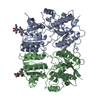
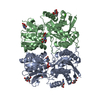
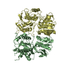
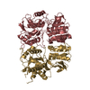
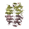
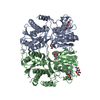
- Assembly
| Deposited unit | 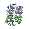
| |||||||||||||||||||||||||||||||||||||||||||||||||||||||||||||||||||||||
|---|---|---|---|---|---|---|---|---|---|---|---|---|---|---|---|---|---|---|---|---|---|---|---|---|---|---|---|---|---|---|---|---|---|---|---|---|---|---|---|---|---|---|---|---|---|---|---|---|---|---|---|---|---|---|---|---|---|---|---|---|---|---|---|---|---|---|---|---|---|---|---|---|
| 1 |
| |||||||||||||||||||||||||||||||||||||||||||||||||||||||||||||||||||||||
| Unit cell |
| |||||||||||||||||||||||||||||||||||||||||||||||||||||||||||||||||||||||
| Noncrystallographic symmetry (NCS) | NCS domain:
NCS domain segments: Ens-ID: 1
|