 Movie
Movie Controller

[English] 日本語
 Yorodumi
- EMDB-9684: Cryo-EM structure of Echovirus 6 complexed with its attachment re... -
+ Open data

- Basic information
| Entry | Database: EMDB / ID: EMD-9684 | |||||||||
|---|---|---|---|---|---|---|---|---|---|---|
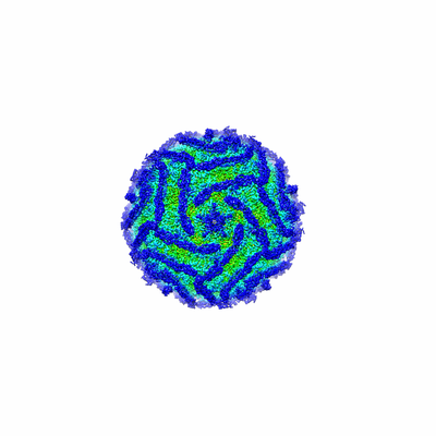
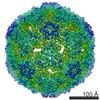
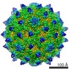
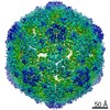
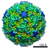
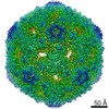
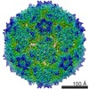
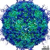
| Title | Cryo-EM structure of Echovirus 6 complexed with its attachment receptor CD55 at PH 5.5 | |||||||||
 Map data Map data | Cryo-EM structure of Echovirus 6 complexed with its attachment receptor CD55 at PH 5.5 | |||||||||
 Sample Sample |
| |||||||||
 Keywords Keywords | Echovirus 6 / CD55 / Cryo-EM / virus-receptor complex / VIRUS | |||||||||
| Function / homology |  Function and homology information Function and homology informationregulation of lipopolysaccharide-mediated signaling pathway / negative regulation of complement activation / regulation of complement-dependent cytotoxicity / regulation of complement activation / respiratory burst / positive regulation of CD4-positive, alpha-beta T cell activation / positive regulation of CD4-positive, alpha-beta T cell proliferation / Class B/2 (Secretin family receptors) / ficolin-1-rich granule membrane / complement activation, classical pathway ...regulation of lipopolysaccharide-mediated signaling pathway / negative regulation of complement activation / regulation of complement-dependent cytotoxicity / regulation of complement activation / respiratory burst / positive regulation of CD4-positive, alpha-beta T cell activation / positive regulation of CD4-positive, alpha-beta T cell proliferation / Class B/2 (Secretin family receptors) / ficolin-1-rich granule membrane / complement activation, classical pathway / side of membrane / transport vesicle / COPI-mediated anterograde transport / endoplasmic reticulum-Golgi intermediate compartment membrane / Regulation of Complement cascade / secretory granule membrane / positive regulation of T cell cytokine production / virus receptor activity / positive regulation of cytosolic calcium ion concentration / membrane raft / Golgi membrane / innate immune response / Neutrophil degranulation / lipid binding / cell surface / extracellular exosome / extracellular region / plasma membrane Similarity search - Function | |||||||||
| Biological species |  Echovirus E6 / Echovirus E6 /  Homo sapiens (human) Homo sapiens (human) | |||||||||
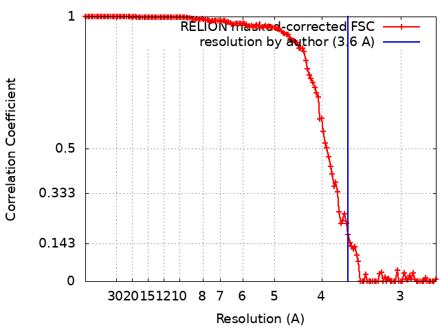
| Method | single particle reconstruction / cryo EM / Resolution: 3.6 Å | |||||||||
 Authors Authors | Gao GF / Liu S | |||||||||
| Funding support |  China, 1 items China, 1 items
| |||||||||
 Citation Citation | Journal: Cell / Year: 2019 Title: Human Neonatal Fc Receptor Is the Cellular Uncoating Receptor for Enterovirus B. Authors: Xin Zhao / Guigen Zhang / Sheng Liu / Xiangpeng Chen / Ruchao Peng / Lianpan Dai / Xiao Qu / Shihua Li / Hao Song / Zhengrong Gao / Pengfei Yuan / Zhiheng Liu / Changyao Li / Zifang Shang / ...Authors: Xin Zhao / Guigen Zhang / Sheng Liu / Xiangpeng Chen / Ruchao Peng / Lianpan Dai / Xiao Qu / Shihua Li / Hao Song / Zhengrong Gao / Pengfei Yuan / Zhiheng Liu / Changyao Li / Zifang Shang / Yan Li / Meifan Zhang / Jianxun Qi / Han Wang / Ning Du / Yan Wu / Yuhai Bi / Shan Gao / Yi Shi / Jinghua Yan / Yong Zhang / Zhengde Xie / Wensheng Wei / George F Gao / Abstract: Enterovirus B (EV-B), a major proportion of the genus Enterovirus in the family Picornaviridae, is the causative agent of severe human infectious diseases. Although cellular receptors for ...Enterovirus B (EV-B), a major proportion of the genus Enterovirus in the family Picornaviridae, is the causative agent of severe human infectious diseases. Although cellular receptors for coxsackievirus B in EV-B have been identified, receptors mediating virus entry, especially the uncoating process of echovirus and other EV-B remain obscure. Here, we found that human neonatal Fc receptor (FcRn) is the uncoating receptor for major EV-B. FcRn binds to the virus particles in the "canyon" through its FCGRT subunit. By obtaining multiple cryo-electron microscopy structures at different stages of virus entry at atomic or near-atomic resolution, we deciphered the underlying mechanisms of enterovirus attachment and uncoating. These structures revealed that different from the attachment receptor CD55, binding of FcRn to the virions induces efficient release of "pocket factor" under acidic conditions and initiates the conformational changes in viral particle, providing a structural basis for understanding the mechanisms of enterovirus entry. | |||||||||
| History |
|
- Structure visualization
| Movie |
Movie viewer |
|---|---|
| Structure viewer | EM map: SurfViewMolmilJmol/JSmol |
| Supplemental images |
- Downloads & links
-EMDB archive
| Map data | emd_9684.map.gz | 41.2 MB | EMDB map data format | |
|---|---|---|---|---|
| Header (meta data) | emd-9684-v30.xmlemd-9684.xml | 20.4 KB 20.4 KB | Display Display | EMDB header |
| FSC (resolution estimation) | emd_9684_fsc.xml | 13.9 KB | Display | FSC data file |
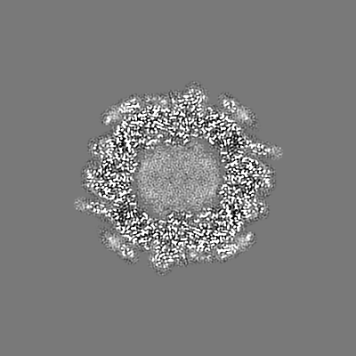
| Images |  emd_9684.png emd_9684.png | 83.2 KB | ||
| Filedesc metadata | emd-9684.cif.gz | 7.4 KB | ||
| Archive directory |  http://ftp.pdbj.org/pub/emdb/structures/EMD-9684ftp://ftp.pdbj.org/pub/emdb/structures/EMD-9684 http://ftp.pdbj.org/pub/emdb/structures/EMD-9684ftp://ftp.pdbj.org/pub/emdb/structures/EMD-9684 | HTTPS FTP |
-Related structure data
| Related structure data |  6iljMC 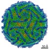 9685C 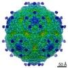 9686C 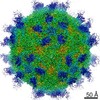 9687C 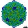 9688C  9689C 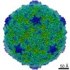 9690C 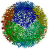 6ilkC 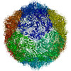 6illC 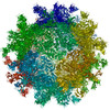 6ilmC 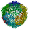 6ilnC 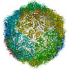 6iloC 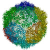 6ilpC M: atomic model generated by this map C: citing same article ( |
|---|---|
| Similar structure data |


-Links
| EMDB pages | EMDB (EBI/PDBe) / EMDataResource |
|---|---|
| Related items in Molecule of the Month |


-Map
| File | Download / File: emd_9684.map.gz / Format: CCP4 / Size: 244.1 MB / Type: IMAGE STORED AS FLOATING POINT NUMBER (4 BYTES) | ||||||||||||||||||||||||||||||||||||||||||||||||||||||||||||
|---|---|---|---|---|---|---|---|---|---|---|---|---|---|---|---|---|---|---|---|---|---|---|---|---|---|---|---|---|---|---|---|---|---|---|---|---|---|---|---|---|---|---|---|---|---|---|---|---|---|---|---|---|---|---|---|---|---|---|---|---|---|
| Annotation | Cryo-EM structure of Echovirus 6 complexed with its attachment receptor CD55 at PH 5.5 | ||||||||||||||||||||||||||||||||||||||||||||||||||||||||||||
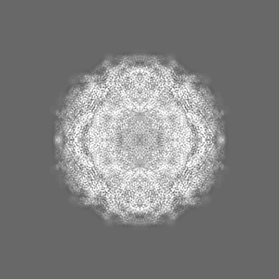
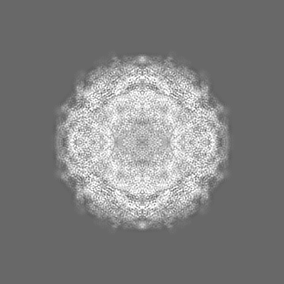
| Projections & slices | Image control
Images are generated by Spider. | ||||||||||||||||||||||||||||||||||||||||||||||||||||||||||||
| Voxel size | X=Y=Z: 1.35 Å | ||||||||||||||||||||||||||||||||||||||||||||||||||||||||||||
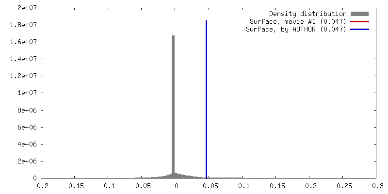
| Density |
| ||||||||||||||||||||||||||||||||||||||||||||||||||||||||||||
| Symmetry | Space group: 1 | ||||||||||||||||||||||||||||||||||||||||||||||||||||||||||||
| Details | EMDB XML:
CCP4 map header:
| ||||||||||||||||||||||||||||||||||||||||||||||||||||||||||||
 Z (Sec.)
 Y (Row.)
 X (Col.)


-Supplemental data
- Sample components
-Entire : Cryo-EM structure of Echovirus 6 complexed with its attachment re...
| Entire | Name: Cryo-EM structure of Echovirus 6 complexed with its attachment receptor CD55 at PH 5.5 |
|---|---|
| Components |
|
-Supramolecule #1: Cryo-EM structure of Echovirus 6 complexed with its attachment re...
| Supramolecule | Name: Cryo-EM structure of Echovirus 6 complexed with its attachment receptor CD55 at PH 5.5 type: complex / ID: 1 / Parent: 0 / Macromolecule list: #1-#5 |
|---|---|
| Source (natural) | Organism: Echovirus E6 |
-Macromolecule #1: Capsid protein VP1
| Macromolecule | Name: Capsid protein VP1 / type: protein_or_peptide / ID: 1 / Number of copies: 1 / Enantiomer: LEVO |
|---|---|
| Source (natural) | Organism: Echovirus E6 |
| Molecular weight | Theoretical: 31.707332 KDa |
| Sequence | String: VVRVADTMPS GPSNSESIPA LTAAETGHTS QVVPSDTIQT RHVRNFHVRS ESSVENFLSR SACVYIVEYK TRDDTPDKMY DSWVINTRQ VAQLRRKLEF FTYVRFDVEV TFVITSVQDD STRQNTDTPA LTHQIMYVPP GGPIPQAVDD YNWQTSTNPS V FWTEGNAP ...String: VVRVADTMPS GPSNSESIPA LTAAETGHTS QVVPSDTIQT RHVRNFHVRS ESSVENFLSR SACVYIVEYK TRDDTPDKMY DSWVINTRQ VAQLRRKLEF FTYVRFDVEV TFVITSVQDD STRQNTDTPA LTHQIMYVPP GGPIPQAVDD YNWQTSTNPS V FWTEGNAP PRMSIPFMSV GNAYSNFYDG WSHFSQTGVY GFNTLNNMGK LYFRHVNDKT ISPITSKVRI YFKPKHVKAW VP RPPRLCE YTHKDNVDFE PKGVTTSRTQ LTISNSTHVE N |
-Macromolecule #2: Capsid protein VP2
| Macromolecule | Name: Capsid protein VP2 / type: protein_or_peptide / ID: 2 / Number of copies: 1 / Enantiomer: LEVO |
|---|---|
| Source (natural) | Organism: Echovirus E6 |
| Molecular weight | Theoretical: 28.06452 KDa |
| Sequence | String: SDRVRSITLG NSTITTQESA NVVVGYGVWP DYLSDEEATA EDQPTQPDVA TCRFYTLDSV SWMKESQGWW WKFPDALRDM GLFGQNMQY HYLGRSGYTI HVQCNASKFH QGCLLVVCVP EAEMGAANIN EKINREHLSN GEVANTFSGT KSSNTNDVQQ A VFNAGMGV ...String: SDRVRSITLG NSTITTQESA NVVVGYGVWP DYLSDEEATA EDQPTQPDVA TCRFYTLDSV SWMKESQGWW WKFPDALRDM GLFGQNMQY HYLGRSGYTI HVQCNASKFH QGCLLVVCVP EAEMGAANIN EKINREHLSN GEVANTFSGT KSSNTNDVQQ A VFNAGMGV AVGNLTIFPH QWINLRTNNC ATIVMPYINS VPMDNMFRHY NFTLMIIPFA KLDYAAGSST YIPITVTVAP MC AEYNGLR LAGHQ |
-Macromolecule #3: Capsid protein VP3
| Macromolecule | Name: Capsid protein VP3 / type: protein_or_peptide / ID: 3 / Number of copies: 1 / Enantiomer: LEVO |
|---|---|
| Source (natural) | Organism: Echovirus E6 |
| Molecular weight | Theoretical: 26.378936 KDa |
| Sequence | String: GLPVMNTPGS NQFLTSDDYQ SPTAMPQFDV TPEMNIPGEV KNLMEIAEVD SVVPVNNVNE NVNSLEAYRI PVHSVTETGA QVFGFTLQP GADTVMERTL LGEILNYYAN WSGSIKLTFM YCGSAMATGK FLLAYSPPGA GVPKNRREAM LGTHIIWDIG L QSSCVLCV ...String: GLPVMNTPGS NQFLTSDDYQ SPTAMPQFDV TPEMNIPGEV KNLMEIAEVD SVVPVNNVNE NVNSLEAYRI PVHSVTETGA QVFGFTLQP GADTVMERTL LGEILNYYAN WSGSIKLTFM YCGSAMATGK FLLAYSPPGA GVPKNRREAM LGTHIIWDIG L QSSCVLCV PWISQTHYRF VSKDIYTDAG FITCWYQTSI VVPAEVQNQS VILCFVSACN DFSVRLLRDS PFVRQTAFYQ |
-Macromolecule #4: Capsid protein VP4
| Macromolecule | Name: Capsid protein VP4 / type: protein_or_peptide / ID: 4 / Number of copies: 1 / Enantiomer: LEVO |
|---|---|
| Source (natural) | Organism: Echovirus E6 |
| Molecular weight | Theoretical: 7.338014 KDa |
| Sequence | String: GAQVSTQKTG AHETSLSASG NSIIHYTNIN YYKDAASNSA NRQDFTQDPG KFTEPVKDIM VKSLPALN |
-Macromolecule #5: Complement decay-accelerating factor
| Macromolecule | Name: Complement decay-accelerating factor / type: protein_or_peptide / ID: 5 / Number of copies: 1 / Enantiomer: LEVO |
|---|---|
| Source (natural) | Organism: Homo sapiens (human) |
| Molecular weight | Theoretical: 21.307959 KDa |
| Recombinant expression | Organism: Homo sapiens (human) |
| Sequence | String: CNRSCEVPTR LNSASLKQPY ITQNYFPVGT VVEYECRPGY RREPSLSPKL TCLQNLKWST AVEFCKKKSC PNPGEIRNGQ IDVPGGILF GATISFSCNT GYKLFGSTSS FCLISGSSVQ WSDPLPECRE IYCPAPPQID NGIIQGERDH YGYRQSVTYA C NKGFTMIG ...String: CNRSCEVPTR LNSASLKQPY ITQNYFPVGT VVEYECRPGY RREPSLSPKL TCLQNLKWST AVEFCKKKSC PNPGEIRNGQ IDVPGGILF GATISFSCNT GYKLFGSTSS FCLISGSSVQ WSDPLPECRE IYCPAPPQID NGIIQGERDH YGYRQSVTYA C NKGFTMIG EHSIYCTVNN DEGEWSGPPP ECRG UniProtKB: Complement decay-accelerating factor |
-Macromolecule #6: SPHINGOSINE
| Macromolecule | Name: SPHINGOSINE / type: ligand / ID: 6 / Number of copies: 1 / Formula: SPH |
|---|---|
| Molecular weight | Theoretical: 299.492 Da |
| Chemical component information |  ChemComp-SPH: |
-Experimental details
-Structure determination
| Method | cryo EM |
|---|---|
 Processing Processing | single particle reconstruction |
| Aggregation state | particle |
-Sample preparation
| Buffer | pH: 5.5 |
|---|---|
| Vitrification | Cryogen name: ETHANE / Chamber humidity: 100 % / Chamber temperature: 277 K / Instrument: FEI VITROBOT MARK IV |
- Electron microscopy
| Microscope | FEI TITAN KRIOS |
|---|---|
| Image recording | Film or detector model: GATAN K2 QUANTUM (4k x 4k) / Detector mode: SUPER-RESOLUTION / Average electron dose: 40.0 e/Å2 |
| Electron beam | Acceleration voltage: 300 kV / Electron source:  FIELD EMISSION GUN FIELD EMISSION GUN |
| Electron optics | Illumination mode: FLOOD BEAM / Imaging mode: BRIGHT FIELD |
| Experimental equipment |  Model: Titan Krios / Image courtesy: FEI Company |
+Image processing
-Atomic model buiding 1
| Refinement | Space: REAL / Protocol: FLEXIBLE FIT |
|---|---|
| Output model | PDB-6ilj: |
