 Movie
Movie Controller


+ Open data

- Basic information
| Entry | Database: EMDB / ID: EMD-6829 | |||||||||
|---|---|---|---|---|---|---|---|---|---|---|
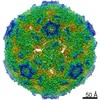
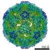
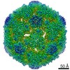
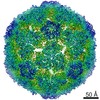
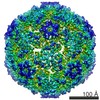
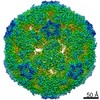
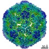
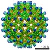
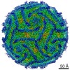
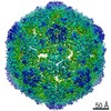
| Title | Cryo-EM Structure of CVA6 VLP | |||||||||
 Map data Map data | ||||||||||
 Sample Sample |
| |||||||||
 Keywords Keywords | Coxsackievirus A6 / virus-like particle / cryo-EM / near-atomic resolution structure / epitope / VIRUS LIKE PARTICLE | |||||||||
| Function / homology |  Function and homology information Function and homology informationsymbiont-mediated suppression of host cytoplasmic pattern recognition receptor signaling pathway via inhibition of MDA-5 activity / picornain 2A / symbiont-mediated suppression of host mRNA export from nucleus / symbiont genome entry into host cell via pore formation in plasma membrane / picornain 3C / T=pseudo3 icosahedral viral capsid / host cell cytoplasmic vesicle membrane / ribonucleoside triphosphate phosphatase activity / nucleoside-triphosphate phosphatase / channel activity ...symbiont-mediated suppression of host cytoplasmic pattern recognition receptor signaling pathway via inhibition of MDA-5 activity / picornain 2A / symbiont-mediated suppression of host mRNA export from nucleus / symbiont genome entry into host cell via pore formation in plasma membrane / picornain 3C / T=pseudo3 icosahedral viral capsid / host cell cytoplasmic vesicle membrane / ribonucleoside triphosphate phosphatase activity / nucleoside-triphosphate phosphatase / channel activity / monoatomic ion transmembrane transport / host cell cytoplasm / DNA replication / RNA helicase activity / endocytosis involved in viral entry into host cell / symbiont-mediated activation of host autophagy / RNA-directed RNA polymerase / cysteine-type endopeptidase activity / viral RNA genome replication / RNA-directed RNA polymerase activity / symbiont entry into host cell / virion attachment to host cell / host cell nucleus / DNA-templated transcription / structural molecule activity / proteolysis / RNA binding / zinc ion binding / ATP binding Similarity search - Function | |||||||||
| Biological species |  Coxsackievirus A6 Coxsackievirus A6 | |||||||||
| Method | single particle reconstruction / cryo EM / Resolution: 3.0 Å | |||||||||
 Authors Authors | Chen J / Zhang C | |||||||||
 Citation Citation | Journal: J Virol / Year: 2018 Title: A 3.0-Angstrom Resolution Cryo-Electron Microscopy Structure and Antigenic Sites of Coxsackievirus A6-Like Particles. Authors: Jinhuan Chen / Chao Zhang / Yu Zhou / Xiang Zhang / Chaoyun Shen / Xiaohua Ye / Wen Jiang / Zhong Huang / Yao Cong /   Abstract: Coxsackievirus A6 (CVA6) has recently emerged as one of the predominant causative agents of hand, foot, and mouth disease (HFMD). The structure of the CVA6 mature viral particle has not been solved ...Coxsackievirus A6 (CVA6) has recently emerged as one of the predominant causative agents of hand, foot, and mouth disease (HFMD). The structure of the CVA6 mature viral particle has not been solved thus far. Our previous work shows that recombinant virus-like particles (VLPs) of CVA6 represent a promising CVA6 vaccine candidate. Here, we report the first cryo-electron microscopy (cryo-EM) structure of the CVA6 VLP at 3.0-Å resolution. The CVA6 VLP exhibits the characteristic features of enteroviruses but presents an open channel at the 2-fold axis and an empty, collapsed VP1 pocket, which is broadly similar to the structures of the enterovirus 71 (EV71) VLP and coxsackievirus A16 (CVA16) 135S expanded particle, indicating that the CVA6 VLP is in an expanded conformation. Structural comparisons reveal that two common salt bridges within protomers are maintained in the CVA6 VLP and other viruses of the genus, implying that these salt bridges may play a critical role in enteroviral protomer assembly. However, there are apparent structural differences among the CVA6 VLP, EV71 VLP, and CVA16 135S particle in the surface-exposed loops and C termini of subunit proteins, which are often antigenic sites for enteroviruses. By immunological assays, we identified two CVA6-specific linear B-cell epitopes (designated P42 and P59) located at the GH loop and the C-terminal region of VP1, respectively, in agreement with the structure-based prediction of antigenic sites. Our findings elucidate the structural basis and important antigenic sites of the CVA6 VLP as a strong vaccine candidate and also provide insight into enteroviral protomer assembly. Coxsackievirus A6 (CVA6) is becoming one of the major pathogens causing hand, foot, and mouth disease (HFMD), leading to significant morbidity and mortality in children and adults. However, no vaccine is currently available to prevent CVA6 infection. Our previous work shows that recombinant virus-like particles (VLPs) of CVA6 are a promising CVA6 vaccine candidate. Here, we present a 3.0-Å structure of the CVA6 VLP determined by cryo-electron microscopy. The overall architecture of the CVA6 VLP is similar to those of the expanded structures of enterovirus 71 (EV71) and coxsackievirus A16 (CVA16), but careful structural comparisons reveal significant differences in the surface-exposed loops and C termini of each capsid protein of these particles. In addition, we identified two CVA6-specific linear B-cell epitopes and mapped them to the GH loop and the C-terminal region of VP1, respectively. Collectively, our findings provide a structural basis and important antigenic information for CVA6 VLP vaccine development. | |||||||||
| History |
|
- Structure visualization
| Movie |
Movie viewer |
|---|---|
| Structure viewer | EM map: SurfViewMolmilJmol/JSmol |
| Supplemental images |

- Downloads & links
-EMDB archive
| Map data | emd_6829.map.gz | 103.7 MB | EMDB map data format | |
|---|---|---|---|---|
| Header (meta data) | emd-6829-v30.xmlemd-6829.xml | 16 KB 16 KB | Display Display | EMDB header |
| Images | 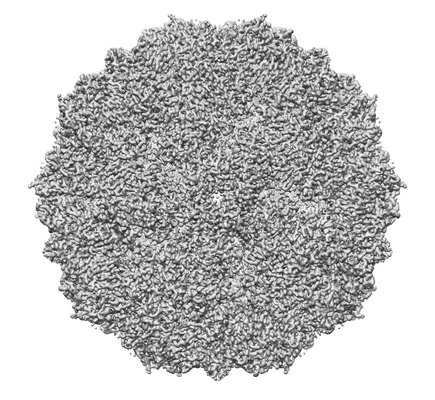 emd_6829.png emd_6829.png | 99.3 KB | ||
| Filedesc metadata | emd-6829.cif.gz | 6.2 KB | ||
| Archive directory |  http://ftp.pdbj.org/pub/emdb/structures/EMD-6829ftp://ftp.pdbj.org/pub/emdb/structures/EMD-6829 http://ftp.pdbj.org/pub/emdb/structures/EMD-6829ftp://ftp.pdbj.org/pub/emdb/structures/EMD-6829 | HTTPS FTP |
-Related structure data
| Related structure data |  5yhqMC M: atomic model generated by this map C: citing same article ( |
|---|---|
| Similar structure data |
-Links
| EMDB pages | EMDB (EBI/PDBe) / EMDataResource |
|---|---|
| Related items in Molecule of the Month |


-Map
| File | Download / File: emd_6829.map.gz / Format: CCP4 / Size: 421.9 MB / Type: IMAGE STORED AS FLOATING POINT NUMBER (4 BYTES) | ||||||||||||||||||||||||||||||||||||||||||||||||||||||||||||
|---|---|---|---|---|---|---|---|---|---|---|---|---|---|---|---|---|---|---|---|---|---|---|---|---|---|---|---|---|---|---|---|---|---|---|---|---|---|---|---|---|---|---|---|---|---|---|---|---|---|---|---|---|---|---|---|---|---|---|---|---|---|
| Projections & slices | Image control
Images are generated by Spider. | ||||||||||||||||||||||||||||||||||||||||||||||||||||||||||||
| Voxel size | X=Y=Z: 0.82 Å | ||||||||||||||||||||||||||||||||||||||||||||||||||||||||||||
| Density |
| ||||||||||||||||||||||||||||||||||||||||||||||||||||||||||||
| Symmetry | Space group: 1 | ||||||||||||||||||||||||||||||||||||||||||||||||||||||||||||
| Details | EMDB XML:
CCP4 map header:
| ||||||||||||||||||||||||||||||||||||||||||||||||||||||||||||
 Z (Sec.)
 Y (Row.)
 X (Col.)


-Supplemental data
- Sample components
-Entire : Coxsackievirus A6
| Entire | Name: Coxsackievirus A6 |
|---|---|
| Components |
|
-Supramolecule #1: Coxsackievirus A6
| Supramolecule | Name: Coxsackievirus A6 / type: virus / ID: 1 / Parent: 0 / Macromolecule list: all / NCBI-ID: 86107 / Sci species name: Coxsackievirus A6 / Virus type: VIRUS-LIKE PARTICLE / Virus isolate: OTHER / Virus enveloped: No / Virus empty: Yes |
|---|
-Macromolecule #1: Capsid protein VP1
| Macromolecule | Name: Capsid protein VP1 / type: protein_or_peptide / ID: 1 / Number of copies: 1 / Enantiomer: LEVO |
|---|---|
| Source (natural) | Organism: Coxsackievirus A6 |
| Molecular weight | Theoretical: 33.644395 KDa |
| Recombinant expression | Organism:   Spodoptera frugiperda (fall armyworm) Spodoptera frugiperda (fall armyworm) |
| Sequence | String: NDPISNAIEN AVSTLADTTI SRVTAANTAA SSHSLGTGRV PALQAAETGA SSNASDENLI ETRCVMNRNG VNEASVEHFY SRAGLVGVV EVKDSGTSQD GYTVWPIDVM GFVQQRRKLE LSTYMRFDAE FTFVSNLNDS TTPGMLLQYM YVPPGAPKPD G RKSYQWQT ...String: NDPISNAIEN AVSTLADTTI SRVTAANTAA SSHSLGTGRV PALQAAETGA SSNASDENLI ETRCVMNRNG VNEASVEHFY SRAGLVGVV EVKDSGTSQD GYTVWPIDVM GFVQQRRKLE LSTYMRFDAE FTFVSNLNDS TTPGMLLQYM YVPPGAPKPD G RKSYQWQT ATNPSIFAKL SDPPPQVSVP FMSPASAYQW FYDGYPTFGE HKQATNLQYG QCPNNMMGHF AIRTVSESTT GK NVHVRVY MRIKHVRAWV PRPFRSQAYM VKNYPTYSQT ISNTAADRAS ITTTDYEGGV PANPQRTF UniProtKB: Genome polyprotein |
-Macromolecule #2: Capsid protein VP3
| Macromolecule | Name: Capsid protein VP3 / type: protein_or_peptide / ID: 2 / Number of copies: 1 / Enantiomer: LEVO |
|---|---|
| Source (natural) | Organism: Coxsackievirus A6 |
| Molecular weight | Theoretical: 26.375834 KDa |
| Recombinant expression | Organism: Spodoptera frugiperda (fall armyworm) |
| Sequence | String: GLPTELKPGT NQFLTTDDGT SPPILPGFEP TPLIHIPGEF TSLLDLCRIE TILEVNNTTG TTGVNRLLIP VRAQNNVDQL CASFQVDPG RNGPWQSTMV GQICRYYTQW SGSLKVTFMF TGSFMATGKM LIAYTPPGSA QPTTREAAML GTHIVWDFGL Q SSVTLVIP ...String: GLPTELKPGT NQFLTTDDGT SPPILPGFEP TPLIHIPGEF TSLLDLCRIE TILEVNNTTG TTGVNRLLIP VRAQNNVDQL CASFQVDPG RNGPWQSTMV GQICRYYTQW SGSLKVTFMF TGSFMATGKM LIAYTPPGSA QPTTREAAML GTHIVWDFGL Q SSVTLVIP WISNTHFRAV KTGGVYDYYA TGIVTIWYQT NFVVPPDTPS EANIIALGAA QENFTLKLCK DTDEIRQTAE YQ UniProtKB: Genome polyprotein |
-Macromolecule #3: capsid protein VP0
| Macromolecule | Name: capsid protein VP0 / type: protein_or_peptide / ID: 3 / Number of copies: 1 / Enantiomer: LEVO |
|---|---|
| Source (natural) | Organism: Coxsackievirus A6 |
| Molecular weight | Theoretical: 35.351426 KDa |
| Recombinant expression | Organism: Spodoptera frugiperda (fall armyworm) |
| Sequence | String: MGAQVSAQKS GTHETGNIAT EGSTINFTNI NYYKDSYAAS ASRQDFTQDP TKFTSPVLDA IKEAAAPLQS PSVEACGYSD RVAQLTVGN STITTQEAAN IVLSYGEWPG YCPSTDATAV DKPTRPDVSV NRFYTLSTKS WKTESTGWYW KFPDVLNDTG V FGQNAQFH ...String: MGAQVSAQKS GTHETGNIAT EGSTINFTNI NYYKDSYAAS ASRQDFTQDP TKFTSPVLDA IKEAAAPLQS PSVEACGYSD RVAQLTVGN STITTQEAAN IVLSYGEWPG YCPSTDATAV DKPTRPDVSV NRFYTLSTKS WKTESTGWYW KFPDVLNDTG V FGQNAQFH YLYRSGFCMH VQCNASKFHQ GALLVVVIPE FVVAASSPAT KPNGQGLYPD FAHTNPGKEG QVFRDPYVLD AG IPLSQAL VFPHQWINLR TNNCATIIMP YVNALPFDSA LNHSNFGLAV IPISPLKYCN GATTEVPITL TIAPLNSEFS GLR QAIKQ UniProtKB: Genome polyprotein |
-Experimental details
-Structure determination
| Method | cryo EM |
|---|---|
 Processing Processing | single particle reconstruction |
| Aggregation state | particle |
-Sample preparation
| Buffer | pH: 7.2 |
|---|---|
| Vitrification | Cryogen name: ETHANE |
- Electron microscopy
| Microscope | FEI TITAN KRIOS |
|---|---|
| Image recording | Film or detector model: GATAN K2 SUMMIT (4k x 4k) / Average electron dose: 42.0 e/Å2 |
| Electron beam | Acceleration voltage: 300 kV / Electron source:  FIELD EMISSION GUN FIELD EMISSION GUN |
| Electron optics | Illumination mode: FLOOD BEAM / Imaging mode: BRIGHT FIELD |
| Experimental equipment |  Model: Titan Krios / Image courtesy: FEI Company |
