 Movie
Movie Controller


+ Open data

- Basic information
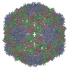
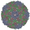
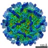
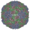
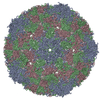
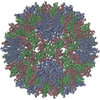
| Entry | Database: PDB / ID: 5yhq | |||||||||||||||||||||||||||||||||
|---|---|---|---|---|---|---|---|---|---|---|---|---|---|---|---|---|---|---|---|---|---|---|---|---|---|---|---|---|---|---|---|---|---|---|
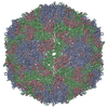
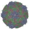
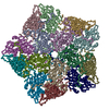
| Title | Cryo-EM Structure of CVA6 VLP | |||||||||||||||||||||||||||||||||
 Components Components |
| |||||||||||||||||||||||||||||||||
 Keywords Keywords | VIRUS LIKE PARTICLE / Coxsackievirus A6 / virus-like particle / cryo-EM / near-atomic resolution structure / epitope | |||||||||||||||||||||||||||||||||
| Function / homology |  Function and homology information Function and homology informationsymbiont-mediated suppression of host cytoplasmic pattern recognition receptor signaling pathway via inhibition of MDA-5 activity / picornain 2A / symbiont-mediated suppression of host mRNA export from nucleus / symbiont genome entry into host cell via pore formation in plasma membrane / picornain 3C / T=pseudo3 icosahedral viral capsid / host cell cytoplasmic vesicle membrane / ribonucleoside triphosphate phosphatase activity / nucleoside-triphosphate phosphatase / channel activity ...symbiont-mediated suppression of host cytoplasmic pattern recognition receptor signaling pathway via inhibition of MDA-5 activity / picornain 2A / symbiont-mediated suppression of host mRNA export from nucleus / symbiont genome entry into host cell via pore formation in plasma membrane / picornain 3C / T=pseudo3 icosahedral viral capsid / host cell cytoplasmic vesicle membrane / ribonucleoside triphosphate phosphatase activity / nucleoside-triphosphate phosphatase / channel activity / monoatomic ion transmembrane transport / host cell cytoplasm / DNA replication / RNA helicase activity / endocytosis involved in viral entry into host cell / symbiont-mediated activation of host autophagy / RNA-directed RNA polymerase / cysteine-type endopeptidase activity / viral RNA genome replication / RNA-directed RNA polymerase activity / symbiont entry into host cell / virion attachment to host cell / host cell nucleus / DNA-templated transcription / structural molecule activity / proteolysis / RNA binding / zinc ion binding / ATP binding Similarity search - Function | |||||||||||||||||||||||||||||||||
| Biological species |  Coxsackievirus A6 Coxsackievirus A6 | |||||||||||||||||||||||||||||||||
| Method | ELECTRON MICROSCOPY / single particle reconstruction / cryo EM / Resolution: 3 Å | |||||||||||||||||||||||||||||||||
 Authors Authors | Chen, J. / Zhang, C. / Huang, Z. / Cong, Y. | |||||||||||||||||||||||||||||||||
 Citation Citation | Journal: J Virol / Year: 2018 Title: A 3.0-Angstrom Resolution Cryo-Electron Microscopy Structure and Antigenic Sites of Coxsackievirus A6-Like Particles. Authors: Jinhuan Chen / Chao Zhang / Yu Zhou / Xiang Zhang / Chaoyun Shen / Xiaohua Ye / Wen Jiang / Zhong Huang / Yao Cong /   Abstract: Coxsackievirus A6 (CVA6) has recently emerged as one of the predominant causative agents of hand, foot, and mouth disease (HFMD). The structure of the CVA6 mature viral particle has not been solved ...Coxsackievirus A6 (CVA6) has recently emerged as one of the predominant causative agents of hand, foot, and mouth disease (HFMD). The structure of the CVA6 mature viral particle has not been solved thus far. Our previous work shows that recombinant virus-like particles (VLPs) of CVA6 represent a promising CVA6 vaccine candidate. Here, we report the first cryo-electron microscopy (cryo-EM) structure of the CVA6 VLP at 3.0-Å resolution. The CVA6 VLP exhibits the characteristic features of enteroviruses but presents an open channel at the 2-fold axis and an empty, collapsed VP1 pocket, which is broadly similar to the structures of the enterovirus 71 (EV71) VLP and coxsackievirus A16 (CVA16) 135S expanded particle, indicating that the CVA6 VLP is in an expanded conformation. Structural comparisons reveal that two common salt bridges within protomers are maintained in the CVA6 VLP and other viruses of the genus, implying that these salt bridges may play a critical role in enteroviral protomer assembly. However, there are apparent structural differences among the CVA6 VLP, EV71 VLP, and CVA16 135S particle in the surface-exposed loops and C termini of subunit proteins, which are often antigenic sites for enteroviruses. By immunological assays, we identified two CVA6-specific linear B-cell epitopes (designated P42 and P59) located at the GH loop and the C-terminal region of VP1, respectively, in agreement with the structure-based prediction of antigenic sites. Our findings elucidate the structural basis and important antigenic sites of the CVA6 VLP as a strong vaccine candidate and also provide insight into enteroviral protomer assembly. Coxsackievirus A6 (CVA6) is becoming one of the major pathogens causing hand, foot, and mouth disease (HFMD), leading to significant morbidity and mortality in children and adults. However, no vaccine is currently available to prevent CVA6 infection. Our previous work shows that recombinant virus-like particles (VLPs) of CVA6 are a promising CVA6 vaccine candidate. Here, we present a 3.0-Å structure of the CVA6 VLP determined by cryo-electron microscopy. The overall architecture of the CVA6 VLP is similar to those of the expanded structures of enterovirus 71 (EV71) and coxsackievirus A16 (CVA16), but careful structural comparisons reveal significant differences in the surface-exposed loops and C termini of each capsid protein of these particles. In addition, we identified two CVA6-specific linear B-cell epitopes and mapped them to the GH loop and the C-terminal region of VP1, respectively. Collectively, our findings provide a structural basis and important antigenic information for CVA6 VLP vaccine development. | |||||||||||||||||||||||||||||||||
| History |
|
- Structure visualization
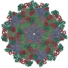
| Movie |
Movie viewer |
|---|---|
| Structure viewer | Molecule: MolmilJmol/JSmol |
- Downloads & links
-Download
| PDBx/mmCIF format | 5yhq.cif.gz | 138.8 KB | Display | PDBx/mmCIF format |
|---|---|---|---|---|
| PDB format | pdb5yhq.ent.gz | 105.3 KB | Display | PDB format |
| PDBx/mmJSON format | 5yhq.json.gz | Tree view | PDBx/mmJSON format | |
| Others |  Other downloads Other downloads |
-Validation report
| Arichive directory | https://data.pdbj.org/pub/pdb/validation_reports/yh/5yhqftp://data.pdbj.org/pub/pdb/validation_reports/yh/5yhq | HTTPS FTP |
|---|
-Related structure data
| Related structure data | 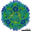 6829MC M: map data used to model this data C: citing same article ( |
|---|---|
| Similar structure data |
-Links
 PDBj
PDBj

- Assembly
| Deposited unit | 
|
|---|---|
| 1 | x 60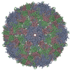
|
| 2 |
|
| 3 | x 5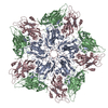
|
| 4 | x 6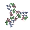
|
| 5 | 
|
| Symmetry | Point symmetry: (Schoenflies symbol: I (icosahedral)) |
-Components
| #1: Protein | Mass: 33644.395 Da / Num. of mol.: 1 Source method: isolated from a genetically manipulated source Source: (gene. exp.) Coxsackievirus A6 / Gene: VP1 / Production host:   Spodoptera frugiperda (fall armyworm) / References: UniProt: Q9YLP0 Spodoptera frugiperda (fall armyworm) / References: UniProt: Q9YLP0 |
|---|---|
| #2: Protein | Mass: 26375.834 Da / Num. of mol.: 1 / Fragment: UNP RESIDUES 326-565 Source method: isolated from a genetically manipulated source Source: (gene. exp.) Coxsackievirus A6 / Production host: Spodoptera frugiperda (fall armyworm) / References: UniProt: Q6JKS2 |
| #3: Protein | Mass: 35351.426 Da / Num. of mol.: 1 / Fragment: UNP RESIDUES 1-325 Source method: isolated from a genetically manipulated source Source: (gene. exp.) Coxsackievirus A6 / Production host: Spodoptera frugiperda (fall armyworm) / References: UniProt: Q6JKS2 |
| Has protein modification | N |
-Experimental details
-Experiment
| Experiment | Method: ELECTRON MICROSCOPY |
|---|---|
| EM experiment | Aggregation state: PARTICLE / 3D reconstruction method: single particle reconstruction |
- Sample preparation
| Component | Name: Coxsackievirus A6 / Type: VIRUS / Entity ID: all / Source: RECOMBINANT |
|---|---|
| Source (natural) | Organism: Coxsackievirus A6 |
| Source (recombinant) | Organism: Spodoptera frugiperda (fall armyworm) |
| Details of virus | Empty: YES / Enveloped: NO / Isolate: OTHER / Type: VIRUS-LIKE PARTICLE |
| Buffer solution | pH: 7.2 |
| Specimen | Embedding applied: NO / Shadowing applied: NO / Staining applied: NO / Vitrification applied: YES |
| Vitrification | Cryogen name: ETHANE |
- Electron microscopy imaging
| Experimental equipment |  Model: Titan Krios / Image courtesy: FEI Company |
|---|---|
| Microscopy | Model: FEI TITAN KRIOS |
| Electron gun | Electron source:  FIELD EMISSION GUN / Accelerating voltage: 300 kV / Illumination mode: FLOOD BEAM FIELD EMISSION GUN / Accelerating voltage: 300 kV / Illumination mode: FLOOD BEAM |
| Electron lens | Mode: BRIGHT FIELD |
| Image recording | Electron dose: 42 e/Å2 / Film or detector model: GATAN K2 SUMMIT (4k x 4k) |
- Processing
| Software | Name: PHENIX / Version: 1.10.1_2155: / Classification: refinement | ||||||||||||||||||||||||
|---|---|---|---|---|---|---|---|---|---|---|---|---|---|---|---|---|---|---|---|---|---|---|---|---|---|
| EM software | Name: PHENIX / Category: model refinement | ||||||||||||||||||||||||
| CTF correction | Type: PHASE FLIPPING AND AMPLITUDE CORRECTION | ||||||||||||||||||||||||
| 3D reconstruction | Resolution: 3 Å / Resolution method: FSC 0.143 CUT-OFF / Num. of particles: 11596 / Symmetry type: POINT | ||||||||||||||||||||||||
| Refinement | Highest resolution: 3 Å | ||||||||||||||||||||||||
| Refine LS restraints |
|
