- EMDB-8646: Locally-refined cryo-EM structure of the small ribosomal subunit ... -
+
データを開く
IDまたはキーワード:
読み込み中...
-
基本情報
登録情報
データベース: EMDB / ID: EMD-8646
タイトル
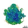
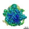
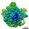
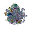
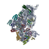
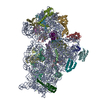
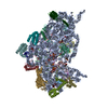
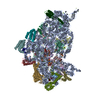
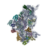
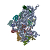
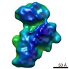
Locally-refined cryo-EM structure of the small ribosomal subunit from Mycobacterium tuberculosis
マップデータ
small ribosomal subunit from Mycobacterium tuberculosis
試料
複合体: Ribosome from human pathogen Mycobacterium tuberculosis
機能・相同性
機能・相同性情報
ribosomal small subunit assembly / ribosome biogenesis / ribosomal small subunit biogenesis / small ribosomal subunit / small ribosomal subunit rRNA binding / cytosolic small ribosomal subunit / tRNA binding / rRNA binding / structural constituent of ribosome / ribosome ...ribosomal small subunit assembly / ribosome biogenesis / ribosomal small subunit biogenesis / small ribosomal subunit / small ribosomal subunit rRNA binding / cytosolic small ribosomal subunit / tRNA binding / rRNA binding / structural constituent of ribosome / ribosome / translation / ribonucleoprotein complex / mRNA binding / RNA binding / zinc ion binding / cytoplasm / cytosol 類似検索 - 分子機能
Ribosomal protein S14, type Z / Ribosomal protein S16, conserved site / Ribosomal protein S16 signature. / Ribosomal protein S6, conserved site / Ribosomal protein S6 signature. / Ribosomal protein S3, bacterial-type / Ribosomal protein S13, bacterial-type / Ribosomal protein S19, bacterial-type / Ribosomal protein S7, bacterial/organellar-type / Ribosomal protein S11, bacterial-type ...Ribosomal protein S14, type Z / Ribosomal protein S16, conserved site / Ribosomal protein S16 signature. / Ribosomal protein S6, conserved site / Ribosomal protein S6 signature. / Ribosomal protein S3, bacterial-type / Ribosomal protein S13, bacterial-type / Ribosomal protein S19, bacterial-type / Ribosomal protein S7, bacterial/organellar-type / Ribosomal protein S11, bacterial-type / Ribosomal protein S20 / Ribosomal protein S20 superfamily / Ribosomal protein S20 / Ribosomal protein S4, bacterial-type / Ribosomal protein S5, bacterial-type / 30S ribosomal protein S17 / Ribosomal protein S6, plastid/chloroplast / Ribosomal protein S14/S29 / Ribosomal protein S18, conserved site / Ribosomal protein S18 signature. / Ribosomal protein S9, bacterial/plastid / Ribosomal protein S16 / Ribosomal protein S16 domain superfamily / Ribosomal protein S16 / Ribosomal protein S15, bacterial-type / Ribosomal protein S6 / Ribosomal protein S6 / Ribosomal protein S6 superfamily / Ribosomal protein S12, bacterial-type / Translation elongation factor EF1B/ribosomal protein S6 / Ribosomal protein S18 / Ribosomal protein S18 / Ribosomal protein S18 superfamily / K Homology domain / K homology RNA-binding domain / Ribosomal protein S3, conserved site / Ribosomal protein S3 signature. / Ribosomal protein S10, conserved site / Ribosomal protein S10 signature. / : / Ribosomal protein S14, conserved site / Ribosomal protein S14 signature. / KH domain / Type-2 KH domain profile. / K Homology domain, type 2 / Ribosomal protein S3, C-terminal / Ribosomal protein S3, C-terminal domain / Ribosomal protein S3, C-terminal domain superfamily / Ribosomal protein S15/S19, conserved site / Ribosomal protein S19 signature. / Ribosomal protein S10 / Ribosomal protein S19/S15 / Ribosomal protein S19/S15, superfamily / Ribosomal protein S19 / Ribosomal protein S5, N-terminal, conserved site / Ribosomal protein S5 signature. / Ribosomal protein S7, conserved site / Ribosomal protein S7 signature. / : / K homology domain superfamily, prokaryotic type / Ribosomal protein S17, conserved site / Ribosomal protein S17 signature. / Ribosomal protein S5 / S5 double stranded RNA-binding domain profile. / Ribosomal protein S5, N-terminal / Ribosomal protein S13, conserved site / Ribosomal protein S13 signature. / Ribosomal protein S5, C-terminal / Ribosomal protein S5, N-terminal domain / Ribosomal protein S13 / 30s ribosomal protein S13, C-terminal / Ribosomal protein S13/S18 / Ribosomal protein S5, C-terminal domain / Ribosomal protein S13 family profile. / Ribosomal protein S4/S9 N-terminal domain / Ribosomal protein S8 signature. / Ribosomal protein S4, conserved site / Ribosomal protein S4 signature. / Ribosomal protein S4/S9 N-terminal domain / Ribosomal protein S4/S9, N-terminal / K homology domain-like, alpha/beta / Ribosomal protein S15 signature. / Ribosomal protein S14 / Ribosomal protein S14p/S29e / Ribosomal protein S4/S9 / Ribosomal protein S8 / Ribosomal protein S8 superfamily / Ribosomal protein S8 / S4 RNA-binding domain profile. / Ribosomal protein S13-like, H2TH / Ribosomal protein S10p/S20e / S4 RNA-binding domain / Ribosomal S11, conserved site / S4 domain / Ribosomal protein S11 signature. / Ribosomal protein S10 domain / Ribosomal protein S10 domain superfamily / Ribosomal protein S10p/S20e / RNA-binding S4 domain / Ribosomal protein S9, conserved site 類似検索 - ドメイン・相同性
Small ribosomal subunit protein uS4 / Small ribosomal subunit protein uS13 / Small ribosomal subunit protein bS20 / Small ribosomal subunit protein uS3 / Small ribosomal subunit protein uS10 / Small ribosomal subunit protein uS14 / Small ribosomal subunit protein uS11 / Small ribosomal subunit protein uS9 / Small ribosomal subunit protein uS8 / Small ribosomal subunit protein uS19 ...Small ribosomal subunit protein uS4 / Small ribosomal subunit protein uS13 / Small ribosomal subunit protein bS20 / Small ribosomal subunit protein uS3 / Small ribosomal subunit protein uS10 / Small ribosomal subunit protein uS14 / Small ribosomal subunit protein uS11 / Small ribosomal subunit protein uS9 / Small ribosomal subunit protein uS8 / Small ribosomal subunit protein uS19 / Small ribosomal subunit protein bS16 / Small ribosomal subunit protein uS15 / Small ribosomal subunit protein uS5 / Small ribosomal subunit protein uS7 / Small ribosomal subunit protein bS6 / Small ribosomal subunit protein bS18 / Small ribosomal subunit protein uS17 / Small ribosomal subunit protein uS12 類似検索 - 構成要素
ジャーナル: Nucleic Acids Res / 年: 2017 タイトル: Structural insights into species-specific features of the ribosome from the human pathogen Mycobacterium tuberculosis. 著者: Kailu Yang / Jeng-Yih Chang / Zhicheng Cui / Xiaojun Li / Ran Meng / Lijun Duan / Jirapat Thongchol / Joanita Jakana / Christoph M Huwe / James C Sacchettini / Junjie Zhang / 要旨: Ribosomes from Mycobacterium tuberculosis (Mtb) possess species-specific ribosomal RNA (rRNA) expansion segments and ribosomal proteins (rProtein). Here, we present the near-atomic structures of the ...Ribosomes from Mycobacterium tuberculosis (Mtb) possess species-specific ribosomal RNA (rRNA) expansion segments and ribosomal proteins (rProtein). Here, we present the near-atomic structures of the Mtb 50S ribosomal subunit and the complete Mtb 70S ribosome, solved by cryo-electron microscopy. Upon joining of the large and small ribosomal subunits, a 100-nt long expansion segment of the Mtb 23S rRNA, named H54a or the 'handle', switches interactions from with rRNA helix H68 and rProtein uL2 to with rProtein bS6, forming a new intersubunit bridge 'B9'. In Mtb 70S, bridge B9 is mostly maintained, leading to correlated motions among the handle, the L1 stalk and the small subunit in the rotated and non-rotated states. Two new protein densities were discovered near the decoding center and the peptidyl transferase center, respectively. These results provide a structural basis for studying translation in Mtb as well as developing new tuberculosis drugs.
 ムービー
ムービー コントローラー


 データを開く
 基本情報
基本情報 マップデータ
 試料
試料 機能・相同性情報
 Mycobacterium tuberculosis (結核菌)
 データ登録者
 引用
引用

 構造の表示
構造の表示
 ダウンロードとリンク
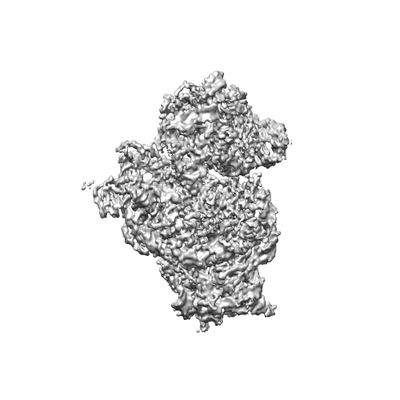 emd_8646.png
 http://ftp.pdbj.org/pub/emdb/structures/EMD-8646
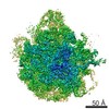


 Z (Sec.)
 Y (Row.)
 X (Col.)


 試料の構成要素
 解析
解析 電子顕微鏡法
 FIELD EMISSION GUN

