 Movie
Movie Controller

[English] 日本語
 Yorodumi
- PDB-5o5j: Structure of the 30S small ribosomal subunit from Mycobacterium s... -
+ Open data

- Basic information
| Entry | Database: PDB / ID: 5o5j | |||||||||
|---|---|---|---|---|---|---|---|---|---|---|
| Title | Structure of the 30S small ribosomal subunit from Mycobacterium smegmatis | |||||||||
 Components Components |
| |||||||||
 Keywords Keywords | RIBOSOME / translation | |||||||||
| Function / homology |  Function and homology information Function and homology informationribosomal small subunit assembly / ribosome biogenesis / ribosomal small subunit biogenesis / small ribosomal subunit / small ribosomal subunit rRNA binding / cytosolic small ribosomal subunit / tRNA binding / rRNA binding / structural constituent of ribosome / ribosome ...ribosomal small subunit assembly / ribosome biogenesis / ribosomal small subunit biogenesis / small ribosomal subunit / small ribosomal subunit rRNA binding / cytosolic small ribosomal subunit / tRNA binding / rRNA binding / structural constituent of ribosome / ribosome / translation / ribonucleoprotein complex / mRNA binding / RNA binding / zinc ion binding / metal ion binding / cytosol / cytoplasm Similarity search - Function | |||||||||
| Biological species |  Mycobacterium smegmatis str. MC2 155 (bacteria) Mycobacterium smegmatis str. MC2 155 (bacteria) | |||||||||
| Method | ELECTRON MICROSCOPY / single particle reconstruction / cryo EM / Resolution: 3.45 Å | |||||||||
 Authors Authors | Hentschel, J. / Burnside, C. / Mignot, I. / Leibundgut, M. / Boehringer, D. / Ban, N. | |||||||||
| Funding support |  Switzerland, 2items Switzerland, 2items
| |||||||||
 Citation Citation | Journal: Cell Rep / Year: 2017 Title: The Complete Structure of the Mycobacterium smegmatis 70S Ribosome. Authors: Jendrik Hentschel / Chloe Burnside / Ingrid Mignot / Marc Leibundgut / Daniel Boehringer / Nenad Ban / Abstract: The ribosome carries out the synthesis of proteins in every living cell. It consequently represents a frontline target in anti-microbial therapy. Tuberculosis ranks among the leading causes of death ...The ribosome carries out the synthesis of proteins in every living cell. It consequently represents a frontline target in anti-microbial therapy. Tuberculosis ranks among the leading causes of death worldwide, due in large part to the combination of difficult-to-treat latency and antibiotic resistance. Here, we present the 3.3-Å cryo-EM structure of the 70S ribosome of Mycobacterium smegmatis, a close relative to the human pathogen Mycobacterium tuberculosis. The structure reveals two additional ribosomal proteins and localizes them to the vicinity of drug-target sites in both the catalytic center and the decoding site of the ribosome. Furthermore, we visualized actinobacterium-specific rRNA and protein expansions that extensively remodel the ribosomal surface with implications for polysome organization. Our results provide a foundation for understanding the idiosyncrasies of mycobacterial translation and reveal atomic details of the structure that will facilitate the design of anti-tubercular therapeutics. | |||||||||
| History |
|
- Structure visualization
| Movie |
Movie viewer |
|---|---|
| Structure viewer | Molecule: MolmilJmol/JSmol |
- Downloads & links
-Download
| PDBx/mmCIF format | 5o5j.cif.gz | 1.3 MB | Display | PDBx/mmCIF format |
|---|---|---|---|---|
| PDB format | pdb5o5j.ent.gz | 1 MB | Display | PDB format |
| PDBx/mmJSON format | 5o5j.json.gz | Tree view | PDBx/mmJSON format | |
| Others |  Other downloads Other downloads |
-Validation report
| Arichive directory | https://data.pdbj.org/pub/pdb/validation_reports/o5/5o5jftp://data.pdbj.org/pub/pdb/validation_reports/o5/5o5j | HTTPS FTP |
|---|
-Related structure data
| Related structure data | 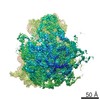 3748MC 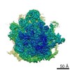 3750C 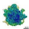 3751C 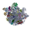 5o60C  5o61C M: map data used to model this data C: citing same article ( |
|---|---|
| Similar structure data |


-Links
 PDBj
PDBj


- Assembly
| Deposited unit | 
|
|---|---|
| 1 |
|
-Components
-RNA chain , 3 types, 3 molecules AWX
| #1: RNA chain | Mass: 495373.656 Da / Num. of mol.: 1 / Source method: isolated from a natural source Source: (natural) Mycobacterium smegmatis str. MC2 155 (bacteria)References: GenBank: 118168627 |
|---|---|
| #22: RNA chain | Mass: 6140.736 Da / Num. of mol.: 1 / Source method: isolated from a natural source Source: (natural) Mycobacterium smegmatis str. MC2 155 (bacteria) |
| #23: RNA chain | Mass: 1791.053 Da / Num. of mol.: 1 / Source method: isolated from a natural source Source: (natural) Mycobacterium smegmatis str. MC2 155 (bacteria) |
-30S ribosomal protein ... , 19 types, 19 molecules CDEFGHIJKLMNOPQRSTV
| #3: Protein | Mass: 30191.227 Da / Num. of mol.: 1 / Source method: isolated from a natural source Source: (natural) Mycobacterium smegmatis str. MC2 155 (bacteria)References: UniProt: A0QSD7 |
|---|---|
| #4: Protein | Mass: 23415.787 Da / Num. of mol.: 1 / Source method: isolated from a natural source Source: (natural) Mycobacterium smegmatis str. MC2 155 (bacteria)References: UniProt: A0QSL7 |
| #5: Protein | Mass: 21946.090 Da / Num. of mol.: 1 / Source method: isolated from a natural source Source: (natural) Mycobacterium smegmatis str. MC2 155 (bacteria)References: UniProt: A0QSG6 |
| #6: Protein | Mass: 10991.637 Da / Num. of mol.: 1 / Source method: isolated from a natural source Source: (natural) Mycobacterium smegmatis str. MC2 155 (bacteria)References: UniProt: A0A0D6J3X3 |
| #7: Protein | Mass: 17660.375 Da / Num. of mol.: 1 / Source method: isolated from a natural source Source: (natural) Mycobacterium smegmatis str. MC2 155 (bacteria)References: UniProt: A0QS97 |
| #8: Protein | Mass: 14492.638 Da / Num. of mol.: 1 / Source method: isolated from a natural source Source: (natural) Mycobacterium smegmatis str. MC2 155 (bacteria)References: UniProt: A0QSG3 |
| #9: Protein | Mass: 16794.365 Da / Num. of mol.: 1 / Source method: isolated from a natural source Source: (natural) Mycobacterium smegmatis str. MC2 155 (bacteria)References: UniProt: A0QSP9 |
| #10: Protein | Mass: 11454.313 Da / Num. of mol.: 1 / Source method: isolated from a natural source Source: (natural) Mycobacterium smegmatis str. MC2 155 (bacteria)References: UniProt: A0QSD0 |
| #11: Protein | Mass: 14671.762 Da / Num. of mol.: 1 / Source method: isolated from a natural source Source: (natural) Mycobacterium smegmatis str. MC2 155 (bacteria)References: UniProt: A0QSL6 |
| #12: Protein | Mass: 13896.366 Da / Num. of mol.: 1 / Source method: isolated from a natural source Source: (natural) Mycobacterium smegmatis str. MC2 155 (bacteria)References: UniProt: A0QS96 |
| #13: Protein | Mass: 14249.619 Da / Num. of mol.: 1 / Source method: isolated from a natural source Source: (natural) Mycobacterium smegmatis str. MC2 155 (bacteria)References: UniProt: A0QSL5 |
| #14: Protein | Mass: 6976.409 Da / Num. of mol.: 1 / Source method: isolated from a natural source Source: (natural) Mycobacterium smegmatis str. MC2 155 (bacteria)References: UniProt: A0QSG2 |
| #15: Protein | Mass: 10368.097 Da / Num. of mol.: 1 / Source method: isolated from a natural source Source: (natural) Mycobacterium smegmatis str. MC2 155 (bacteria)References: UniProt: A0QVQ3 |
| #16: Protein | Mass: 16795.207 Da / Num. of mol.: 1 / Source method: isolated from a natural source Source: (natural) Mycobacterium smegmatis str. MC2 155 (bacteria)References: UniProt: A0QV37 |
| #17: Protein | Mass: 11127.002 Da / Num. of mol.: 1 / Source method: isolated from a natural source Source: (natural) Mycobacterium smegmatis str. MC2 155 (bacteria)References: UniProt: A0QSE0 |
| #18: Protein | Mass: 9524.188 Da / Num. of mol.: 1 / Source method: isolated from a natural source Source: (natural) Mycobacterium smegmatis str. MC2 155 (bacteria)References: UniProt: A0R7F7 |
| #19: Protein | Mass: 10800.602 Da / Num. of mol.: 1 / Source method: isolated from a natural source Source: (natural) Mycobacterium smegmatis str. MC2 155 (bacteria)References: UniProt: A0QSD5 |
| #20: Protein | Mass: 9556.104 Da / Num. of mol.: 1 / Source method: isolated from a natural source Source: (natural) Mycobacterium smegmatis str. MC2 155 (bacteria)References: UniProt: A0R102 |
| #21: Protein | Mass: 30145.230 Da / Num. of mol.: 1 / Source method: isolated from a natural source Source: (natural) Mycobacterium smegmatis str. MC2 155 (bacteria)References: UniProt: A0QVB8 |
-Protein/peptide / Protein , 2 types, 2 molecules Bg
| #24: Protein | Mass: 8312.485 Da / Num. of mol.: 1 / Source method: isolated from a natural source Source: (natural) Mycobacterium smegmatis str. MC2 155 (bacteria)References: UniProt: A0R215 |
|---|---|
| #2: Protein/peptide | Mass: 4164.300 Da / Num. of mol.: 1 / Source method: isolated from a natural source Source: (natural) Mycobacterium smegmatis str. MC2 155 (bacteria)References: UniProt: A0QR10 |
-Non-polymers , 2 types, 219 molecules 

| #25: Chemical | ChemComp-MG /  Mass: 24.305 Da / Num. of mol.: 217 / Source method: obtained synthetically / Formula: Mg Mass: 24.305 Da / Num. of mol.: 217 / Source method: obtained synthetically / Formula: Mg#26: Chemical |  Mass: 65.409 Da / Num. of mol.: 2 / Source method: obtained synthetically / Formula: Zn Mass: 65.409 Da / Num. of mol.: 2 / Source method: obtained synthetically / Formula: Zn |
|---|
-Experimental details
-Experiment
| Experiment | Method: ELECTRON MICROSCOPY |
|---|---|
| EM experiment | Aggregation state: PARTICLE / 3D reconstruction method: single particle reconstruction |
- Sample preparation
| Component | Name: 30S small ribosomal subunit / Type: RIBOSOME / Entity ID: #1-#24 / Source: NATURAL |
|---|---|
| Source (natural) | Organism: Mycobacterium smegmatis str. MC2 155 (bacteria) |
| Buffer solution | pH: 7.5 |
| Specimen | Embedding applied: NO / Shadowing applied: NO / Staining applied: NO / Vitrification applied: YES |
| Specimen support | Grid material: COPPER / Grid type: Quantifoil R2/2 |
| Vitrification | Instrument: FEI VITROBOT MARK I / Cryogen name: ETHANE-PROPANE / Humidity: 96 % |
- Electron microscopy imaging
| Experimental equipment |  Model: Titan Krios / Image courtesy: FEI Company |
|---|---|
| Microscopy | Model: FEI TITAN KRIOS |
| Electron gun | Electron source:  FIELD EMISSION GUN / Accelerating voltage: 300 kV / Illumination mode: FLOOD BEAM FIELD EMISSION GUN / Accelerating voltage: 300 kV / Illumination mode: FLOOD BEAM |
| Electron lens | Mode: BRIGHT FIELD / Calibrated magnification: 100719 X |
| Specimen holder | Cryogen: NITROGEN / Specimen holder model: FEI TITAN KRIOS AUTOGRID HOLDER |
| Image recording | Electron dose: 20 e/Å2 / Detector mode: INTEGRATING / Film or detector model: FEI FALCON II (4k x 4k) / Details: FEI EPU data collection |
| Image scans | Movie frames/image: 7 |
- Processing
| Software | Name: PHENIX / Version: 1.9_1692 / Classification: refinement | |||||||||||||||||||||||||||||||||||||||||||||||||||||||||||||||||||||||||||||||||||||||||||||||||||||||||||||||||||||||||||||||||||||||||||||||||||||||||||||||||||||||||||||||||||||||||||||||||||||||||||||||||||||||||
|---|---|---|---|---|---|---|---|---|---|---|---|---|---|---|---|---|---|---|---|---|---|---|---|---|---|---|---|---|---|---|---|---|---|---|---|---|---|---|---|---|---|---|---|---|---|---|---|---|---|---|---|---|---|---|---|---|---|---|---|---|---|---|---|---|---|---|---|---|---|---|---|---|---|---|---|---|---|---|---|---|---|---|---|---|---|---|---|---|---|---|---|---|---|---|---|---|---|---|---|---|---|---|---|---|---|---|---|---|---|---|---|---|---|---|---|---|---|---|---|---|---|---|---|---|---|---|---|---|---|---|---|---|---|---|---|---|---|---|---|---|---|---|---|---|---|---|---|---|---|---|---|---|---|---|---|---|---|---|---|---|---|---|---|---|---|---|---|---|---|---|---|---|---|---|---|---|---|---|---|---|---|---|---|---|---|---|---|---|---|---|---|---|---|---|---|---|---|---|---|---|---|---|---|---|---|---|---|---|---|---|---|---|---|---|---|---|---|---|
| EM software |
| |||||||||||||||||||||||||||||||||||||||||||||||||||||||||||||||||||||||||||||||||||||||||||||||||||||||||||||||||||||||||||||||||||||||||||||||||||||||||||||||||||||||||||||||||||||||||||||||||||||||||||||||||||||||||
| CTF correction | Details: CTF correction in Relion / Type: NONE | |||||||||||||||||||||||||||||||||||||||||||||||||||||||||||||||||||||||||||||||||||||||||||||||||||||||||||||||||||||||||||||||||||||||||||||||||||||||||||||||||||||||||||||||||||||||||||||||||||||||||||||||||||||||||
| Symmetry | Point symmetry: C1 (asymmetric) | |||||||||||||||||||||||||||||||||||||||||||||||||||||||||||||||||||||||||||||||||||||||||||||||||||||||||||||||||||||||||||||||||||||||||||||||||||||||||||||||||||||||||||||||||||||||||||||||||||||||||||||||||||||||||
| 3D reconstruction | Resolution: 3.45 Å / Resolution method: FSC 0.143 CUT-OFF / Num. of particles: 224584 / Symmetry type: POINT | |||||||||||||||||||||||||||||||||||||||||||||||||||||||||||||||||||||||||||||||||||||||||||||||||||||||||||||||||||||||||||||||||||||||||||||||||||||||||||||||||||||||||||||||||||||||||||||||||||||||||||||||||||||||||
| Atomic model building | Protocol: OTHER | |||||||||||||||||||||||||||||||||||||||||||||||||||||||||||||||||||||||||||||||||||||||||||||||||||||||||||||||||||||||||||||||||||||||||||||||||||||||||||||||||||||||||||||||||||||||||||||||||||||||||||||||||||||||||
| Refinement | Resolution: 3.451→49.359 Å / SU ML: 0.45 / σ(F): 0.98 / Phase error: 25.38 / Stereochemistry target values: MLHL
| |||||||||||||||||||||||||||||||||||||||||||||||||||||||||||||||||||||||||||||||||||||||||||||||||||||||||||||||||||||||||||||||||||||||||||||||||||||||||||||||||||||||||||||||||||||||||||||||||||||||||||||||||||||||||
| Solvent computation | Shrinkage radii: 0.9 Å / VDW probe radii: 1.11 Å / Solvent model: FLAT BULK SOLVENT MODEL | |||||||||||||||||||||||||||||||||||||||||||||||||||||||||||||||||||||||||||||||||||||||||||||||||||||||||||||||||||||||||||||||||||||||||||||||||||||||||||||||||||||||||||||||||||||||||||||||||||||||||||||||||||||||||
| Refine LS restraints |
| |||||||||||||||||||||||||||||||||||||||||||||||||||||||||||||||||||||||||||||||||||||||||||||||||||||||||||||||||||||||||||||||||||||||||||||||||||||||||||||||||||||||||||||||||||||||||||||||||||||||||||||||||||||||||
| LS refinement shell |
|
