 ムービー
ムービー コントローラー


+ データを開く

- 基本情報
基本情報
| 登録情報 | データベース: EMDB / ID: EMD-7079 | |||||||||||||||
|---|---|---|---|---|---|---|---|---|---|---|---|---|---|---|---|---|
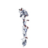
| タイトル | Structure of the 30 kDa HIV-1 RNA Dimerization Signal by a Hybrid Cryo-EM, NMR, and Molecular Dynamics Approach | |||||||||||||||
 マップデータ マップデータ | DIS dimer 3D map from data set 1 (JEOL2200FS microscope, DE12 detector) | |||||||||||||||
 試料 試料 | 30 kDa HIV-1 RNA Dimerization Signal != Human immunodeficiency virus 1 30 kDa HIV-1 RNA Dimerization Signal
| |||||||||||||||
| 生物種 |   Human immunodeficiency virus 1 (ヒト免疫不全ウイルス) Human immunodeficiency virus 1 (ヒト免疫不全ウイルス) | |||||||||||||||
| 手法 | 単粒子再構成法 / クライオ電子顕微鏡法 / 解像度: 9.0 Å | |||||||||||||||
 データ登録者 データ登録者 | Zhang K / Keane S / Su Z / Case D / Ludtke S / Summers M / Chiu W | |||||||||||||||
| 資金援助 |  米国, 4件 米国, 4件
| |||||||||||||||
 引用 引用 | ジャーナル: Structure / 年: 2018 タイトル: Structure of the 30 kDa HIV-1 RNA Dimerization Signal by a Hybrid Cryo-EM, NMR, and Molecular Dynamics Approach. 著者: Kaiming Zhang / Sarah C Keane / Zhaoming Su / Rossitza N Irobalieva / Muyuan Chen / Verna Van / Carly A Sciandra / Jan Marchant / Xiao Heng / Michael F Schmid / David A Case / Steven J Ludtke ...著者: Kaiming Zhang / Sarah C Keane / Zhaoming Su / Rossitza N Irobalieva / Muyuan Chen / Verna Van / Carly A Sciandra / Jan Marchant / Xiao Heng / Michael F Schmid / David A Case / Steven J Ludtke / Michael F Summers / Wah Chiu / 要旨: Cryoelectron microscopy (cryo-EM) and nuclear magnetic resonance (NMR) spectroscopy are routinely used to determine structures of macromolecules with molecular weights over 65 and under 25 kDa, ...Cryoelectron microscopy (cryo-EM) and nuclear magnetic resonance (NMR) spectroscopy are routinely used to determine structures of macromolecules with molecular weights over 65 and under 25 kDa, respectively. We combined these techniques to study a 30 kDa HIV-1 dimer initiation site RNA ([DIS]; 47 nt/strand). A 9 Å cryo-EM map clearly shows major groove features of the double helix and a right-handed superhelical twist. Simulated cryo-EM maps generated from time-averaged molecular dynamics trajectories (10 ns) exhibited levels of detail similar to those in the experimental maps, suggesting internal structural flexibility limits the cryo-EM resolution. Simultaneous inclusion of the cryo-EM map and H-edited NMR-derived distance restraints during structure refinement generates a structure consistent with both datasets and supporting a flipped-out base within a conserved purine-rich bulge. Our findings demonstrate the power of combining global and local structural information from these techniques for structure determination of modest-sized RNAs. | |||||||||||||||
| 履歴 |
|
- 構造の表示
構造の表示
| ムービー |
 ムービービューア ムービービューア |
|---|---|
| 構造ビューア | EMマップ: SurfViewMolmilJmol/JSmol |
| 添付画像 |

- ダウンロードとリンク
-EMDBアーカイブ
| マップデータ | emd_7079.map.gz | 7.4 MB | EMDBマップデータ形式 | |
|---|---|---|---|---|
| ヘッダ (付随情報) | emd-7079-v30.xmlemd-7079.xml | 11.7 KB 11.7 KB | 表示 表示 | EMDBヘッダ |
| 画像 | 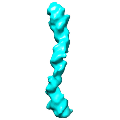 emd_7079.png emd_7079.png | 49 KB | ||
| アーカイブディレクトリ |  http://ftp.pdbj.org/pub/emdb/structures/EMD-7079ftp://ftp.pdbj.org/pub/emdb/structures/EMD-7079 http://ftp.pdbj.org/pub/emdb/structures/EMD-7079ftp://ftp.pdbj.org/pub/emdb/structures/EMD-7079 | HTTPS FTP |
-関連構造データ


-リンク
| EMDBのページ | EMDB (EBI/PDBe) / EMDataResource |
|---|---|
| 「今月の分子」の関連する項目 |

-マップ
| ファイル | ダウンロード / ファイル: emd_7079.map.gz / 形式: CCP4 / 大きさ: 8 MB / タイプ: IMAGE STORED AS FLOATING POINT NUMBER (4 BYTES) | ||||||||||||||||||||||||||||||||||||||||||||||||||||||||||||||||||||
|---|---|---|---|---|---|---|---|---|---|---|---|---|---|---|---|---|---|---|---|---|---|---|---|---|---|---|---|---|---|---|---|---|---|---|---|---|---|---|---|---|---|---|---|---|---|---|---|---|---|---|---|---|---|---|---|---|---|---|---|---|---|---|---|---|---|---|---|---|---|
| 注釈 | DIS dimer 3D map from data set 1 (JEOL2200FS microscope, DE12 detector) | ||||||||||||||||||||||||||||||||||||||||||||||||||||||||||||||||||||
| 投影像・断面図 | 画像のコントロール
画像は Spider により作成 | ||||||||||||||||||||||||||||||||||||||||||||||||||||||||||||||||||||
| ボクセルのサイズ | X=Y=Z: 2.01 Å | ||||||||||||||||||||||||||||||||||||||||||||||||||||||||||||||||||||
| 密度 |
| ||||||||||||||||||||||||||||||||||||||||||||||||||||||||||||||||||||
| 対称性 | 空間群: 1 | ||||||||||||||||||||||||||||||||||||||||||||||||||||||||||||||||||||
| 詳細 | EMDB XML:
CCP4マップ ヘッダ情報:
| ||||||||||||||||||||||||||||||||||||||||||||||||||||||||||||||||||||
 Z (Sec.)
 Y (Row.)
 X (Col.)


-添付データ
- 試料の構成要素
-全体 : 30 kDa HIV-1 RNA Dimerization Signal
| 全体 | 名称: 30 kDa HIV-1 RNA Dimerization Signal |
|---|---|
| 要素 |
|
-超分子 #1: Human immunodeficiency virus 1
| 超分子 | 名称: Human immunodeficiency virus 1 / タイプ: virus / ID: 1 / 親要素: 0 / NCBI-ID: 11676 / 生物種: Human immunodeficiency virus 1 / ウイルスタイプ: VIRION / ウイルス・単離状態: OTHER / ウイルス・エンベロープ: Yes / ウイルス・中空状態: Yes |
|---|---|
| Host system | 生物種: Human immunodeficiency virus 1 (ヒト免疫不全ウイルス) |
| 分子量 | 実験値: 30 KDa |
-実験情報
-構造解析
| 手法 | クライオ電子顕微鏡法 |
|---|---|
 解析 解析 | 単粒子再構成法 |
| 試料の集合状態 | particle |
-試料調製
| 緩衝液 | pH: 8 |
|---|---|
| グリッド | 前処理 - タイプ: GLOW DISCHARGE / 詳細: unspecified |
| 凍結 | 凍結剤: ETHANE / チャンバー内湿度: 100 % / 装置: FEI VITROBOT MARK IV |
| 詳細 | HIV-1 RNA Dimerization Signal |
- 電子顕微鏡法
| 顕微鏡 | JEOL 2200FS |
|---|---|
| 撮影 | フィルム・検出器のモデル: DIRECT ELECTRON DE-12 (4k x 3k) 撮影したグリッド数: 4 / 実像数: 540 / 平均電子線量: 75.0 e/Å2 |
| 電子線 | 加速電圧: 200 kV / 電子線源:  FIELD EMISSION GUN FIELD EMISSION GUN |
| 電子光学系 | 照射モード: FLOOD BEAM / 撮影モード: BRIGHT FIELD / 倍率(公称値): 25000 |
| 試料ステージ | 試料ホルダーモデル: GATAN 626 SINGLE TILT LIQUID NITROGEN CRYO TRANSFER HOLDER ホルダー冷却材: NITROGEN |
