 Movie
Movie Controller

[English] 日本語
 Yorodumi
- PDB-6bg9: HYBRID NMR/CRYO-EM STRUCTURE OF THE HIV-1 RNA DIMERIZATION SIGNAL -
+ Open data

- Basic information
| Entry | Database: PDB / ID: 6bg9 | |||||||||
|---|---|---|---|---|---|---|---|---|---|---|
| Title | HYBRID NMR/CRYO-EM STRUCTURE OF THE HIV-1 RNA DIMERIZATION SIGNAL | |||||||||
 Components Components | RNA dimerization signal | |||||||||
 Keywords Keywords | RNA / RNA INERNAL LOOPS / SHEARED GA PAIRS / GU WOBBLE / S-TURN THERMODYNAMICS | |||||||||
| Function / homology | RNA / RNA (> 10) Function and homology information Function and homology information | |||||||||
| Biological species |  Homo sapiens (human) Homo sapiens (human) | |||||||||
| Method | ELECTRON MICROSCOPY / SOLUTION NMR / subtomogram averaging / molecular dynamics / cryo EM / Resolution: 9 Å | |||||||||
 Authors Authors | Summers, M.F. | |||||||||
| Funding support |  United States, 2items United States, 2items
| |||||||||
 Citation Citation | Journal: Structure / Year: 2018 Title: Structure of the 30 kDa HIV-1 RNA Dimerization Signal by a Hybrid Cryo-EM, NMR, and Molecular Dynamics Approach. Authors: Kaiming Zhang / Sarah C Keane / Zhaoming Su / Rossitza N Irobalieva / Muyuan Chen / Verna Van / Carly A Sciandra / Jan Marchant / Xiao Heng / Michael F Schmid / David A Case / Steven J ...Authors: Kaiming Zhang / Sarah C Keane / Zhaoming Su / Rossitza N Irobalieva / Muyuan Chen / Verna Van / Carly A Sciandra / Jan Marchant / Xiao Heng / Michael F Schmid / David A Case / Steven J Ludtke / Michael F Summers / Wah Chiu / Abstract: Cryoelectron microscopy (cryo-EM) and nuclear magnetic resonance (NMR) spectroscopy are routinely used to determine structures of macromolecules with molecular weights over 65 and under 25 kDa, ...Cryoelectron microscopy (cryo-EM) and nuclear magnetic resonance (NMR) spectroscopy are routinely used to determine structures of macromolecules with molecular weights over 65 and under 25 kDa, respectively. We combined these techniques to study a 30 kDa HIV-1 dimer initiation site RNA ([DIS]; 47 nt/strand). A 9 Å cryo-EM map clearly shows major groove features of the double helix and a right-handed superhelical twist. Simulated cryo-EM maps generated from time-averaged molecular dynamics trajectories (10 ns) exhibited levels of detail similar to those in the experimental maps, suggesting internal structural flexibility limits the cryo-EM resolution. Simultaneous inclusion of the cryo-EM map and H-edited NMR-derived distance restraints during structure refinement generates a structure consistent with both datasets and supporting a flipped-out base within a conserved purine-rich bulge. Our findings demonstrate the power of combining global and local structural information from these techniques for structure determination of modest-sized RNAs. | |||||||||
| History |
|
- Structure visualization
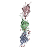
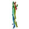
| Movie |
Movie viewer |
|---|---|
| Structure viewer | Molecule: MolmilJmol/JSmol |
- Downloads & links
-Download
| PDBx/mmCIF format | 6bg9.cif.gz | 1.2 MB | Display | PDBx/mmCIF format |
|---|---|---|---|---|
| PDB format | pdb6bg9.ent.gz | 994.7 KB | Display | PDB format |
| PDBx/mmJSON format | 6bg9.json.gz | Tree view | PDBx/mmJSON format | |
| Others |  Other downloads Other downloads |
-Validation report
| Arichive directory | https://data.pdbj.org/pub/pdb/validation_reports/bg/6bg9ftp://data.pdbj.org/pub/pdb/validation_reports/bg/6bg9 | HTTPS FTP |
|---|
-Related structure data
| Related structure data |  7080MC 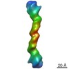 7079C M: map data used to model this data C: citing same article ( |
|---|---|
| Similar structure data | |
| Other databases |


-Links
 PDBj
PDBj


- Assembly
| Deposited unit | 
| |||||||||
|---|---|---|---|---|---|---|---|---|---|---|
| 1 |
| |||||||||
| NMR ensembles |
|
-Components
| #1: RNA chain | Mass: 15359.241 Da / Num. of mol.: 2 Source method: isolated from a genetically manipulated source Source: (gene. exp.) Homo sapiens (human)Production host: in vitro transcription vector pT7-TP(deltai) (others) |
|---|
-Experimental details
-Experiment
| Experiment |
| ||||||||||||||||||
|---|---|---|---|---|---|---|---|---|---|---|---|---|---|---|---|---|---|---|---|
| EM experiment | Aggregation state: PARTICLE / 3D reconstruction method: subtomogram averaging | ||||||||||||||||||
| NMR experiment |
| ||||||||||||||||||
| NMR details | Text: NMR studies conducted with multiple samples prepared by differential nucleotide-specific 2H labeling. |
- Sample preparation
| Component | Name: Human immunodeficiency virus 1 / Type: ORGANELLE OR CELLULAR COMPONENT Details: RNA prepared by in vitro transcription using T7 RNA polymerase Entity ID: all / Source: MULTIPLE SOURCES | ||||||||||||||||||||||||||||||||||||||||||||||||||||||||||||||||||||
|---|---|---|---|---|---|---|---|---|---|---|---|---|---|---|---|---|---|---|---|---|---|---|---|---|---|---|---|---|---|---|---|---|---|---|---|---|---|---|---|---|---|---|---|---|---|---|---|---|---|---|---|---|---|---|---|---|---|---|---|---|---|---|---|---|---|---|---|---|---|
| Molecular weight | Experimental value: NO | ||||||||||||||||||||||||||||||||||||||||||||||||||||||||||||||||||||
| Source (natural) | Organism: Homo sapiens (human) | ||||||||||||||||||||||||||||||||||||||||||||||||||||||||||||||||||||
| Source (recombinant) | Organism: in vitro transcription vector pT7-TP(deltai) (others) | ||||||||||||||||||||||||||||||||||||||||||||||||||||||||||||||||||||
| Details of virus | Isolate: OTHER / Type: VIRUS-LIKE PARTICLE | ||||||||||||||||||||||||||||||||||||||||||||||||||||||||||||||||||||
| Buffer solution | pH: 7.2 | ||||||||||||||||||||||||||||||||||||||||||||||||||||||||||||||||||||
| Specimen | Embedding applied: NO / Shadowing applied: NO / Staining applied: NO / Vitrification applied: YES | ||||||||||||||||||||||||||||||||||||||||||||||||||||||||||||||||||||
| Specimen support | Details: unspecified | ||||||||||||||||||||||||||||||||||||||||||||||||||||||||||||||||||||
| Vitrification | Instrument: FEI VITROBOT MARK IV / Cryogen name: ETHANE / Humidity: 100 % | ||||||||||||||||||||||||||||||||||||||||||||||||||||||||||||||||||||
| Details |
| ||||||||||||||||||||||||||||||||||||||||||||||||||||||||||||||||||||
| Sample |
| ||||||||||||||||||||||||||||||||||||||||||||||||||||||||||||||||||||
| Sample conditions | Details: Samples in D2O, 5 mM NaCl, 140 mM KCl, 1 mM MgCl2, Tris, pH 7.2, T= 308K Ionic strength: 150 mM / Ionic strength err: 10 / Label: All samples / pH: 7.1 pD / Pressure: 1 atm / Temperature: 308 K |
-Data collection
| Microscopy | Model: JEOL 2200FS |
|---|---|
| Electron gun | Electron source:  FIELD EMISSION GUN / Accelerating voltage: 200 kV / Illumination mode: FLOOD BEAM FIELD EMISSION GUN / Accelerating voltage: 200 kV / Illumination mode: FLOOD BEAM |
| Electron lens | Mode: BRIGHT FIELD / Nominal magnification: 25000 X |
| Specimen holder | Cryogen: NITROGEN Specimen holder model: GATAN 626 SINGLE TILT LIQUID NITROGEN CRYO TRANSFER HOLDER |
| Image recording | Electron dose: 75 e/Å2 / Film or detector model: DIRECT ELECTRON DE-12 (4k x 3k) / Num. of grids imaged: 4 |
| NMR spectrometer | Type: Bruker AVANCE / Manufacturer: Bruker / Model: AVANCE / Field strength: 600 MHz |
- Processing
| EM software |
| |||||||||||||||||||||
|---|---|---|---|---|---|---|---|---|---|---|---|---|---|---|---|---|---|---|---|---|---|---|
| CTF correction | Type: PHASE FLIPPING AND AMPLITUDE CORRECTION | |||||||||||||||||||||
| Symmetry | Point symmetry: C2 (2 fold cyclic) | |||||||||||||||||||||
| 3D reconstruction | Resolution: 9 Å / Resolution method: FSC 0.143 CUT-OFF / Num. of particles: 20366 / Symmetry type: POINT | |||||||||||||||||||||
| EM volume selection | Num. of tomograms: 540 / Num. of volumes extracted: 20366 | |||||||||||||||||||||
| NMR software |
| |||||||||||||||||||||
| Refinement | Method: molecular dynamics / Software ordinal: 6 Details: Used GB for solvent simulation, NOEs and EM data simultaneously as restraints | |||||||||||||||||||||
| NMR representative | Selection criteria: lowest energy | |||||||||||||||||||||
| NMR ensemble | Conformer selection criteria: all calculated structures submitted Conformers calculated total number: 20 / Conformers submitted total number: 20 |
