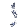
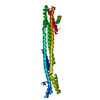
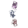
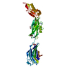
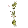
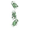
Entry Database : PDB / ID : 5zo1Title Crystal structure of mouse nectin-like molecule 4 (mNecl-4) full ectodomain (Ig1-Ig3), 2.2A Cell adhesion molecule 4 Keywords / / / / / / / / / / / / / Function / homology Function Domain/homology Component
/ / / / / / / / / / / / / / / / / / / / / / / / / / / / / / / / / / / / / / / / / / / / / / / / / / Biological species Mus musculus (house mouse)Method / / / Resolution : 2.201 Å Authors Liu, X. / An, T. / Li, D. / Fan, Z. / Xiang, P. / Li, C. / Ju, W. / Li, J. / Hu, G. / Qin, B. ...Liu, X. / An, T. / Li, D. / Fan, Z. / Xiang, P. / Li, C. / Ju, W. / Li, J. / Hu, G. / Qin, B. / Yin, B. / Wojdyla, J.A. / Wang, M. / Yuan, J. / Qiang, B. / Shu, P. / Cui, S. / Peng, X. Funding support Organization Grant number Country National Key Research and Development Program 2016YFA0100702, 2016YFC092502 National Key Basic Research Program 2013CB531304
Journal : Proc. Natl. Acad. Sci. U.S.A. / Year : 2019Title : Structure of the heterophilic interaction between the nectin-like 4 and nectin-like 1 molecules.Authors : Liu, X. / An, T. / Li, D. / Fan, Z. / Xiang, P. / Li, C. / Ju, W. / Li, J. / Hu, G. / Qin, B. / Yin, B. / Wojdyla, J.A. / Wang, M. / Yuan, J. / Qiang, B. / Shu, P. / Cui, S. / Peng, X. History Deposition Apr 12, 2018 Deposition site / Processing site Revision 1.0 Jan 30, 2019 Provider / Type Revision 1.1 Feb 6, 2019 Group / Database references / Category / citation_authorItem _citation.journal_abbrev / _citation.journal_id_CSD ... _citation.journal_abbrev / _citation.journal_id_CSD / _citation.pdbx_database_id_PubMed / _citation.title / _citation_author.identifier_ORCID Revision 1.2 Feb 20, 2019 Group / Database references / Category / citation_authorItem _citation.journal_volume / _citation.page_first ... _citation.journal_volume / _citation.page_first / _citation.page_last / _citation_author.identifier_ORCID Revision 2.0 Jul 29, 2020 Group Atomic model / Data collection ... Atomic model / Data collection / Derived calculations / Structure summary Category atom_site / chem_comp ... atom_site / chem_comp / entity / pdbx_branch_scheme / pdbx_chem_comp_identifier / pdbx_entity_branch / pdbx_entity_branch_descriptor / pdbx_entity_branch_link / pdbx_entity_branch_list / pdbx_entity_nonpoly / pdbx_nonpoly_scheme / pdbx_struct_assembly_gen / pdbx_struct_special_symmetry / struct_asym / struct_conn / struct_site / struct_site_gen Item _atom_site.B_iso_or_equiv / _atom_site.Cartn_x ... _atom_site.B_iso_or_equiv / _atom_site.Cartn_x / _atom_site.Cartn_y / _atom_site.Cartn_z / _atom_site.auth_asym_id / _atom_site.auth_atom_id / _atom_site.auth_comp_id / _atom_site.auth_seq_id / _atom_site.label_asym_id / _atom_site.label_atom_id / _atom_site.label_comp_id / _atom_site.label_entity_id / _atom_site.type_symbol / _chem_comp.name / _chem_comp.type / _pdbx_struct_assembly_gen.asym_id_list / _pdbx_struct_special_symmetry.label_asym_id / _struct_conn.pdbx_dist_value / _struct_conn.pdbx_leaving_atom_flag / _struct_conn.pdbx_role / _struct_conn.ptnr1_auth_asym_id / _struct_conn.ptnr1_auth_comp_id / _struct_conn.ptnr1_auth_seq_id / _struct_conn.ptnr1_label_asym_id / _struct_conn.ptnr1_label_atom_id / _struct_conn.ptnr1_label_comp_id / _struct_conn.ptnr2_auth_asym_id / _struct_conn.ptnr2_auth_comp_id / _struct_conn.ptnr2_auth_seq_id / _struct_conn.ptnr2_label_asym_id / _struct_conn.ptnr2_label_atom_id / _struct_conn.ptnr2_label_comp_id Description / Provider / Type Revision 2.1 Nov 6, 2024 Group Data collection / Database references ... Data collection / Database references / Derived calculations / Structure summary Category chem_comp / chem_comp_atom ... chem_comp / chem_comp_atom / chem_comp_bond / database_2 / pdbx_entry_details / pdbx_modification_feature / struct_conn Item _chem_comp.pdbx_synonyms / _database_2.pdbx_DOI ... _chem_comp.pdbx_synonyms / _database_2.pdbx_DOI / _database_2.pdbx_database_accession / _struct_conn.pdbx_leaving_atom_flag
Show all Show less
 Movie
Movie Controller

 Yorodumi
 Open data
 Basic information
 Components
 Keywords
 Function and homology information
 X-RAY DIFFRACTION /
  Authors
 China, 2items
  Citation
 Structure visualization
 Downloads & links
 Other downloads
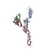


 PDBj
PDBj


 Assembly
 Trichoplusia ni (cabbage looper) / References: UniProt: Q8R464
 Mass: 92.094 Da / Num. of mol.: 2 / Source method: obtained synthetically / Formula: C3H8O3
 Mass: 18.015 Da / Num. of mol.: 188 / Source method: isolated from a natural source / Formula: H2O
 Sample preparation
 / Beamline: X06DA / Wavelength: 1 Å
 Processing